Tympanic part (Latin: pars tympanica ossis temporalis)
Also called the tympanic plate
· It is the part of the temporal bone that surrounds the external acoustic meatus.
· It is a thin, curved plate of bone, shaped like a half-tube and roughly four-sided, that forms the front wall, the floor and part of the back wall of the bony external acoustic meatus.
- Squamous part : the part that occupies the middle and the whole upper part of the bone; it has two surfaces, temporal and cerebral.
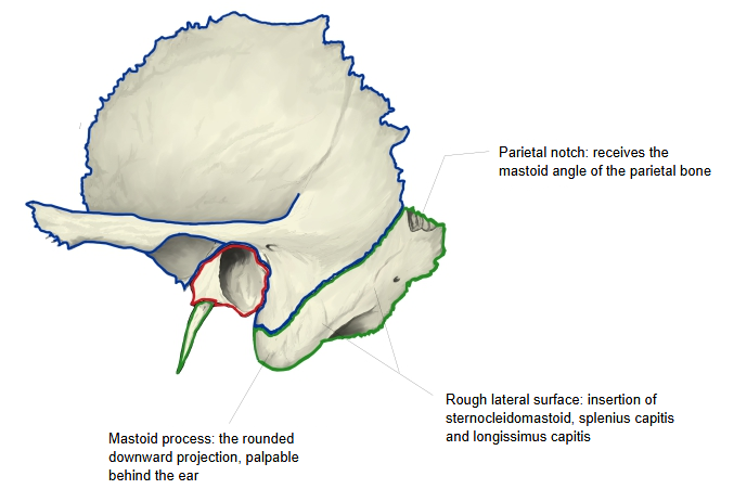
- Petrous part : the lower part, made of two parts: the mastoid part (from the mastoid process up to the parietal notch) and the pyramid (part of the cranial base).
- Tympanic part : the smallest part: a thin, half-tube-shaped plate that forms the front and lower walls of the external acoustic meatus.
· The name comes from the Greek "tympanon", a drum, and refers to the eardrum (tympanic membrane). It does not mean that this part contains the whole tympanic cavity, most of which lies in the petrous part.
· Its surfaces and borders are described separately below.
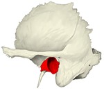 Tympanic part (lateral view)
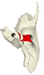 Tympanic part (inferior view)
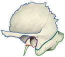 Parts of the temporal bone (outlined)
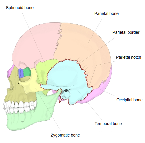
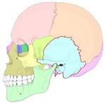 Skull (lateral view), bones coloured
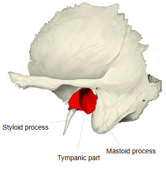
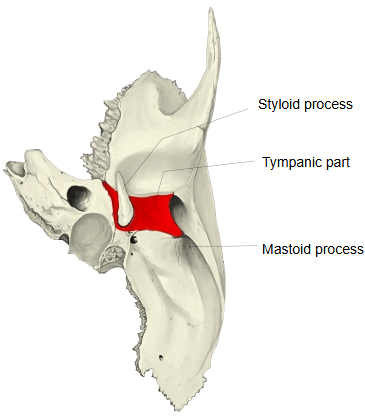
The tympanic part is a small, thin plate that forms the front and lower wall of the external acoustic meatus. It first develops as a separate membrane bone, the tympanic ring, which is still an incomplete ring at birth, so the bony meatus of a newborn is very short; it grows outwards into a plate in early childhood and fuses with the under side of the pyramid. Its upper front edge meets the petrous part at the petrotympanic fissure behind the mandibular fossa (the socket of the jaw joint). Its lower edge forms a sharp ridge on the under side of the pyramid, which continues backwards and outwards as a sheath over the root of the styloid process.
Reference websites
[Wikipedia]
· Tympanic part of the temporal bone, Wikipedia.
· Temporal bone, Wikipedia.
[Websites with illustrations]
· The temporal bone (TeachMeAnatomy)
· Temporal bone: anatomy, parts, sutures and foramina (Kenhub)
· Left temporal bone, outer surface (Gray's Anatomy, fig. 137) (Wikimedia Commons)

· Left temporal bone, inferior surface (Gray's Anatomy, fig. 141) (Wikimedia Commons)

Surfaces
Posterosuperior surface: concave; it lines the bony external acoustic meatus, forming its front wall, floor and part of its back wall. At its inner end a narrow groove, the tympanic sulcus, holds the rim of the tympanic membrane. Anteroinferior surface: four-sided and slightly concave; it forms the back wall of the mandibular fossa and touches the parotid gland.
Borders
Lateral border: free and rough; the cartilaginous part of the external acoustic meatus is attached to it. Medially the tympanic part is fused with the petrous part, below and lateral to the opening of the auditory tube. Upper border: laterally it fuses with the back of the postglenoid process; medially it forms the petrotympanic fissure with the petrous part, through which pass the chorda tympani and the anterior tympanic artery. Behind, it meets the mastoid part at the tympanomastoid fissure, through which the auricular branch of the vagus nerve passes. Lower border: thin and sharp medially; laterally it splits to form a sheath around the root of the styloid process (the vaginal process).
Where it is found
Related terms
References and sources
- Further reading: Tympanic part of the temporal bone, Wikipedia.
- Further reading: Temporal bone, Wikipedia.
- Federative International Programme for Anatomical Terminology (FIPAT). Terminologia Anatomica, 2nd ed. (TA2), 2019.