Latin: glandula submandibularis
Overview
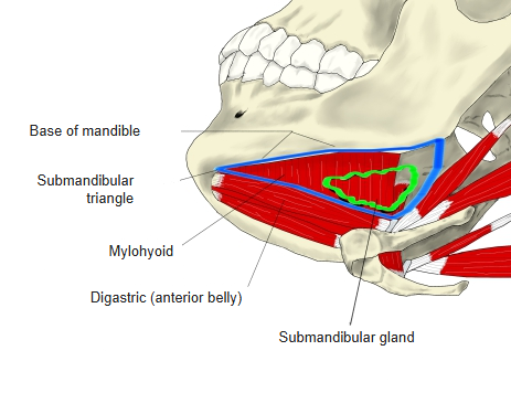
- A major salivary gland in the submandibular triangle, below the mylohyoid, between the base of the mandible and the anterior and posterior bellies of the digastric.
- Flattened oval, about 10–15 g; a mixed gland, mostly serous.
- Produces about two thirds of resting saliva; its share falls when stimulation increases parotid secretion.
- Has a large superficial part and a small deep part, continuous around the back edge of the mylohyoid; enclosed by the investing layer of the deep cervical fascia.
- Its intercalated and striated ducts are much better developed than in the parotid and sublingual glands.
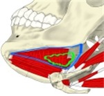
Submandibular triangle 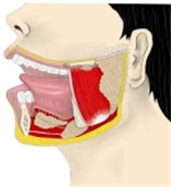
Major salivary glands
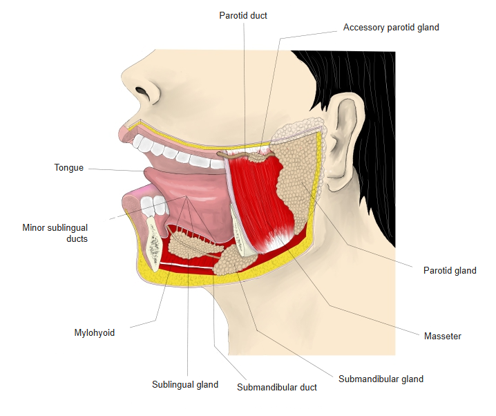
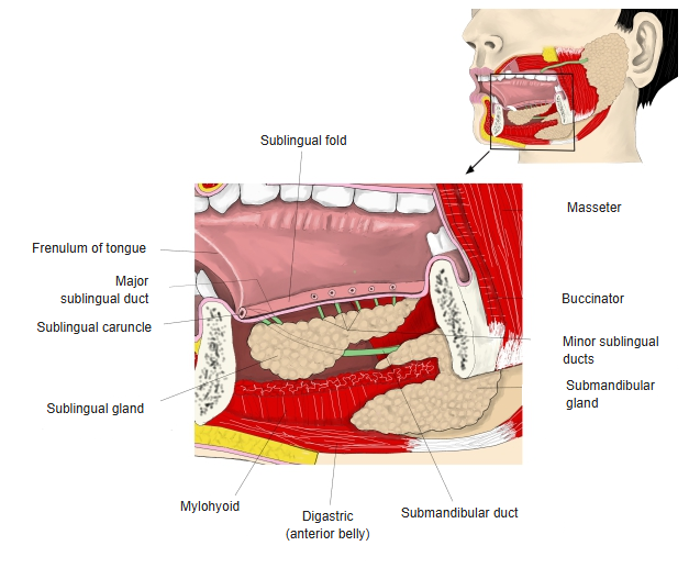
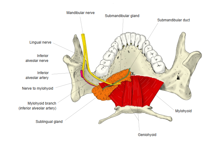
Submandibular duct
- Also called Wharton’s duct; about 5 cm long.
- Leaves the deep part of the gland and runs forward.
- Hooks around the posterior border of the mylohyoid onto its upper surface.
- Is crossed by the lingual nerve, which loops under it from lateral to medial.
- Runs forward along the medial side of the sublingual gland.
- Opens at the sublingual caruncle beside the frenulum of the tongue, alone or together with the major sublingual duct.
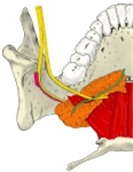
Course of the submandibular duct 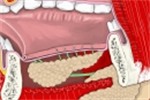
Floor of the mouth from inside 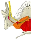
Floor of the mouth from above
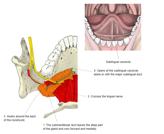
Blood supply, lymph and nerves
| Supply | Source |
|---|---|
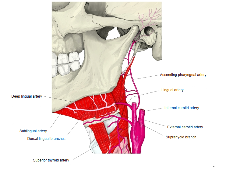
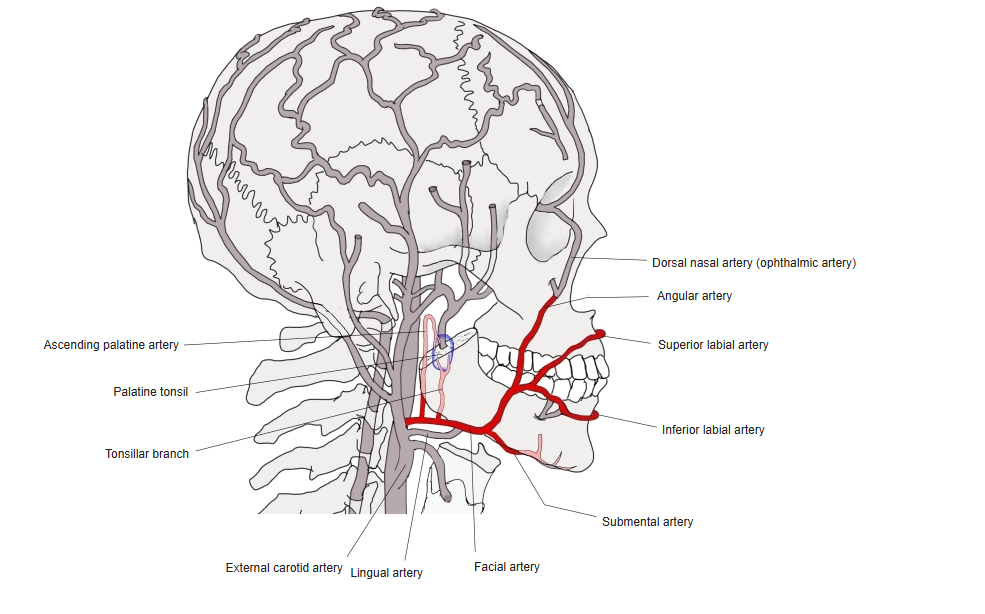
| Arteries | Branches of the facial and lingual arteries |
| Veins | Follow the arteries |
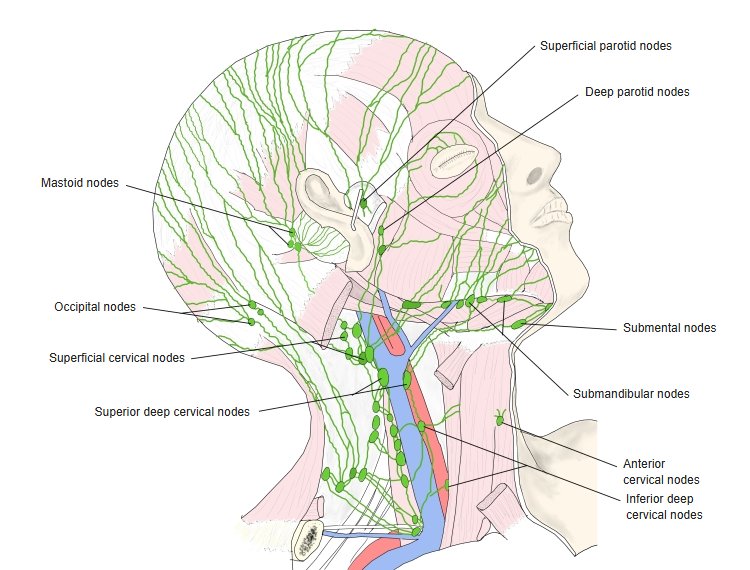
| Lymph | Submandibular nodes |
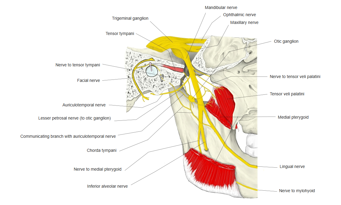
| Parasympathetic (secretomotor) | Facial nerve → chorda tympani → lingual nerve → submandibular ganglion → gland |
| Sympathetic | Superior cervical ganglion, via the plexus around the external carotid and facial arteries |
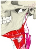
Lingual artery 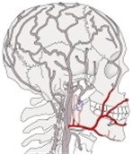
Facial artery 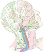
Lymph nodes of the head and neck 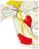
Mandibular nerve (medial view)
References and sources
- Further reading: Submandibular gland, Wikipedia.