Auditory ossicles (Latin: ossicula auditus)
Also called the ear ossicles or the middle ear bones
· They are the smallest bones in the body and are covered by a thin mucous membrane, continuous with the lining of the tympanic cavity.
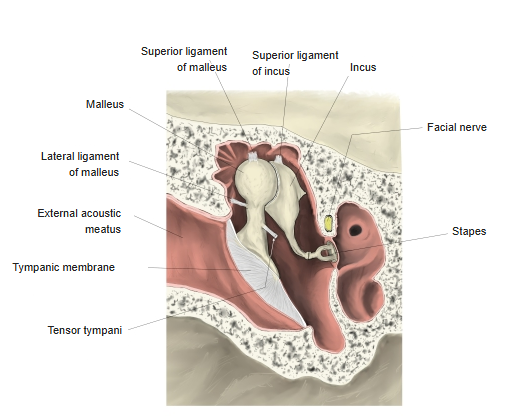
· They are held to the walls of the cavity by small ligaments, for example the malleus by the lateral ligament of malleus and the stapes by the anular ligament of stapes.
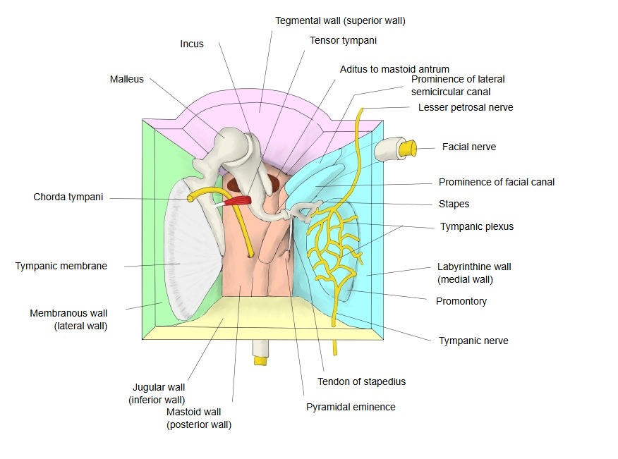
· The chorda tympani, a branch of the facial nerve carrying fibres for taste and for the secretion of saliva, crosses the cavity between the malleus and the incus.
ossicles
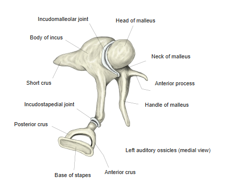
- Malleus : the largest and most lateral ossicle, attached to the tympanic membrane by its handle and lateral process. Three parts (head, neck and handle) and two processes (anterior and lateral). Its head forms the incudomalleolar joint with the incus.
- Incus : lies between the malleus and the stapes; a body and two limbs (short limb and long limb). The tip of the long limb (the lenticular process) forms the incudostapedial joint with the stapes.
- Stapes : the smallest and most medial ossicle: head, anterior and posterior limbs and base (footplate). The base closes the oval window and is attached to its rim by the anular ligament.
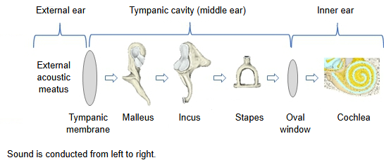
· They carry the vibrations of the eardrum to the fluid of the inner ear and increase their pressure on the way. Two small muscles damp the chain: the tensor tympani on the malleus and the stapedius on the stapes.
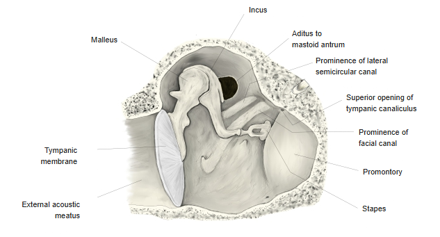
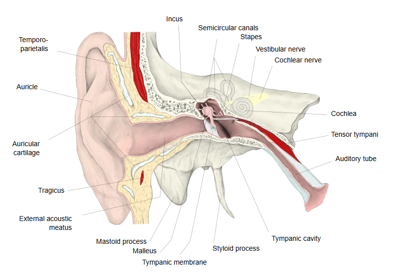
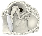 Right tympanic cavity (coronal section)
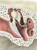 Right tympanic cavity (coronal section)
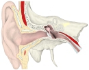 External ear to middle ear
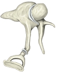 Left auditory ossicles (medial view)
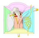 Right tympanic cavity (model)
Reference websites
[Wikipedia]
· Ossicles, Wikipedia.
[Websites with illustrations]
· Ossicles: anatomy and functions (Kenhub)
· The middle ear (TeachMeAnatomy)
· Chain of ossicles and their ligaments (Gray's Anatomy) (Wikimedia Commons)

Where it is found
Related terms
References and sources
- Further reading: Ossicles, Wikipedia.
- Federative International Programme for Anatomical Terminology (FIPAT). Terminologia Anatomica, 2nd ed. (TA2), 2019.