Clivus (Latin: clivus)
- Reference websites
- (Wikipedia, websites with illustrations)
- Structure
- Where it is found
- Related terms
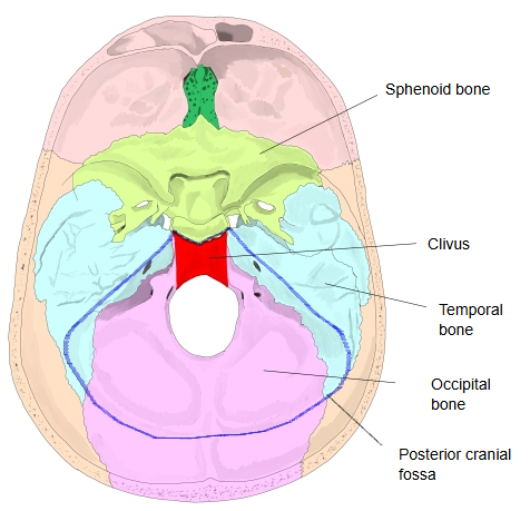
· It forms the most anterior, median part of the posterior cranial fossa, with the petrous parts of the temporal bones on each side.
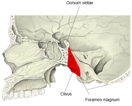
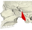 Skull (sagittal section)
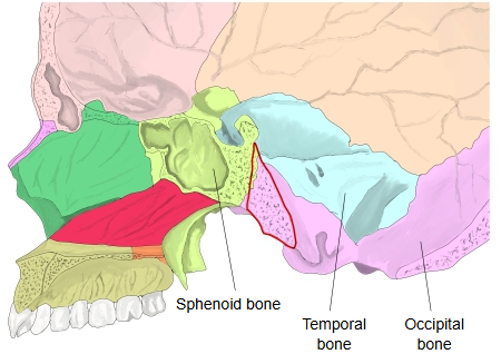
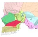 Skull (sagittal section, bones coloured)
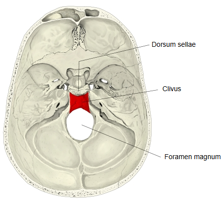
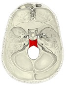 Cranial base (internal view)
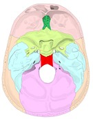 Cranial base (bones coloured)
Reference websites
[Wikipedia]
· Clivus (anatomy), Wikipedia.
· Basilar part of occipital bone, Wikipedia.
[Websites with illustrations]
· The posterior cranial fossa (TeachMeAnatomy)
· The occipital bone (TeachMeAnatomy)
Structure
The clivus is the shallow, sloping surface behind the dorsum sellae; it runs down from the sphenoid bone to the front edge of the foramen magnum. The sphenoidal sinus lies in front of its upper part, and its outer (lower) surface faces the roof of the nasopharynx. On its inner surface lie the pons and the medulla oblongata, with the basilar artery between them and the bone, and the abducens nerve runs up along it. The tectorial membrane and the apical ligament of dens attach near its lower end. The sphenoid and occipital parts are joined by the spheno-occipital synchondrosis, which fuses in adolescence; defects in this fusion can leave small variant pits or canals in the clivus, and a small bony tubercle may form at its lower end.
Where it is found
Related terms
- Occipital bone
- Sphenoid bone
- Foramen magnum
- Sella turcica
- Sphenoidal sinus
- Occipital condyle
- Hypoglossal canal
References and sources
- Further reading: Clivus (anatomy), Wikipedia.
- Further reading: Basilar part of occipital bone, Wikipedia.
- Federative International Programme for Anatomical Terminology (FIPAT). Terminologia Anatomica, 2nd ed. (TA2), 2019.