Sphenoidal sinus (Latin: sinus sphenoidalis)
Also called the sphenoid sinus
- Superior surface : faces the cranial cavity and forms the central part of the cranial fossae. Neighbours: the ethmoid bone in front; behind, the dorsum sellae forms the clivus with the basilar part of the occipital bone. Features: sella turcica, hypophysial fossa, tuberculum sellae, dorsum sellae, middle clinoid process, posterior clinoid process, chiasmatic sulcus, carotid sulcus, lingula of the sphenoid.
- Inferior surface : forms the roof of the pharynx. Neighbour: the vomer.
- Anterior surface : faces the nasal cavity. Neighbour: the ethmoid bone. Features: sphenoidal crest (continuing below into the sphenoidal rostrum), sphenoidal conchae (with the aperture of the sphenoidal sinus).
- Posterior surface : a rough, four-sided surface. Neighbour: the occipital bone.
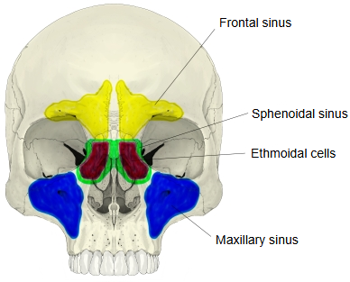
- Maxillary sinus : in the maxilla; the largest of the paranasal sinuses.
- Frontal sinus : in the frontal bone.
- Ethmoidal sinus : in the ethmoid bone, divided into many small spaces (ethmoidal cells).
- Sphenoidal sinus : in the sphenoid bone, behind and deep to the ethmoidal sinus.
· Its average size is about 2.2 cm high, 2 cm wide and 2.2 cm deep.
The nerve supply above follows the description in Wikipedia.
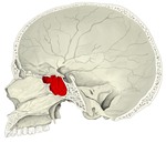 Skull (sagittal section)
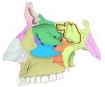 Bones of the nasal cavity (lateral wall)
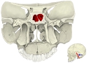 Coronal section through the sinuses
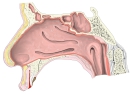 Lateral wall of the nasal cavity
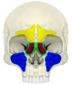 Paranasal sinuses
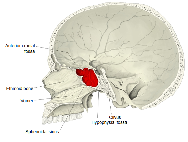
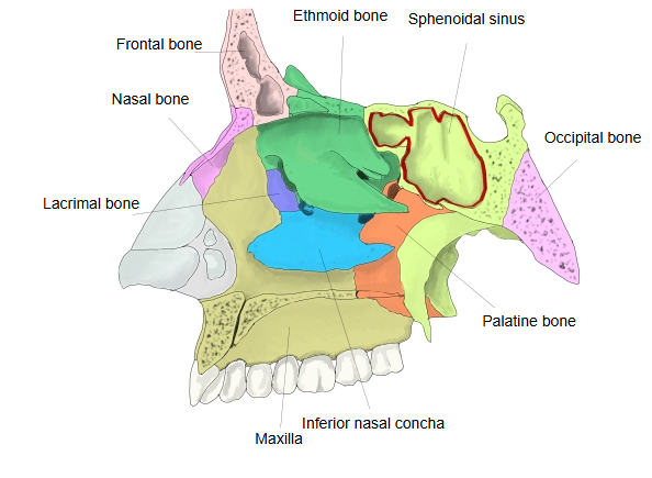
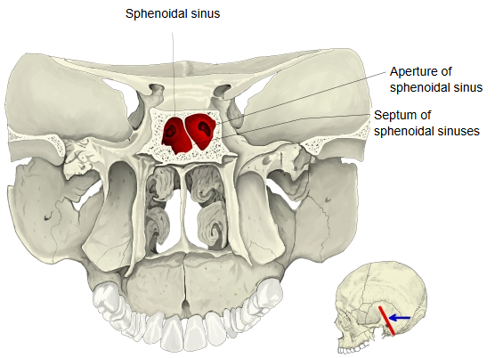
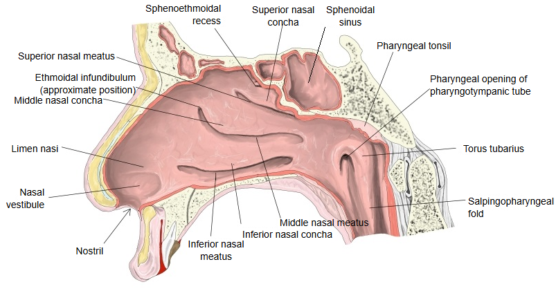
Most of the inside of the body of the sphenoid is taken up by the sphenoidal sinus. Its size varies, and it sometimes extends into the base of the occipital bone that joins the back of the body. It is the paranasal sinus at the skull base. Its pneumatization varies from person to person. The optic canal with the optic nerve and the internal carotid artery lie close to it; seen from inside the sinus, the bony bulges of the carotid artery (carotid prominence) and of the optic canal can be seen, with a hollow between them called the opticocarotid recess.
Because it lies just below the pituitary gland in the sella turcica, the sinus is the usual route for transsphenoidal surgery on the pituitary.
Reference websites
[Wikipedia]
· Sphenoid sinus, Wikipedia.
· Paranasal sinuses, Wikipedia.
· Sphenoid bone, Wikipedia.
[Websites with illustrations]
· The paranasal sinuses (TeachMeAnatomy)
· Lateral wall of nasal cavity (Gray's Anatomy) (Wikimedia Commons)

Where it is found
Related terms
- Body of sphenoid
- Sella turcica
- Sphenoid bone
- Frontal sinus
- Maxillary sinus
- Ethmoidal air cells
- Nasal cavity
References and sources
- Further reading: Sphenoid sinus, Wikipedia.
- Further reading: Paranasal sinuses, Wikipedia.
- Federative International Programme for Anatomical Terminology (FIPAT). Terminologia Anatomica, 2nd ed. (TA2), 2019.