Hypophysial fossa (Latin: fossa hypophysialis)
Also called the pituitary fossa
- Superior surface : faces the cranial cavity and forms the central part of the cranial fossae. Joins: the ethmoid bone in front; behind, the dorsum sellae forms the clivus with the basilar part of the occipital bone.Features: sella turcica, hypophysial fossa, tuberculum sellae, dorsum sellae, middle clinoid process, posterior clinoid process, chiasmatic groove, carotid sulcus, sphenoidal lingula
- Inferior surface : forms the roof of the pharynx. Joins: the vomer (ala of vomer).
- Anterior surface : faces the nasal cavity. Joins: the ethmoid bone (and the orbital processes of the palatine bones).Features: sphenoidal crest (continuing below as the sphenoidal rostrum), sphenoidal concha (aperture of sphenoidal sinus)
- Posterior surface : a rough, four-sided surface. Joins: the occipital bone (basilar part).
| 1 | Superior surface | Faces the cranial cavity and forms the central part of the cranial fossae. Joins: in front the ethmoid bone (cribriform plate). Behind, the back of the dorsum sellae forms the clivus together with the basilar part of the occipital bone. In the middle of the upper surface of the body is a hollow, the sella turcica; in and around it lie the hypophysial fossa, the tuberculum sellae, the middle clinoid process, the chiasmatic groove, the dorsum sellae, the posterior clinoid process, the carotid sulcus and the sphenoidal lingula. |
| 2 | Inferior surface | Forms the roof of the pharynx. Joins: the vomer (ala of vomer). |
| 3 | Anterior surface | Faces the nasal cavity. Joins: the ethmoid bone (and the orbital processes of the palatine bones). Features: the sphenoidal crest, continuing onto the under side as the sphenoidal rostrum, and the sphenoidal concha (with the aperture of the sphenoidal sinus). |
| 4 | Posterior surface | A rough, four-sided surface. Joins: the occipital bone (basilar part). |
Note: on this site the sella turcica is taken to consist of the following.
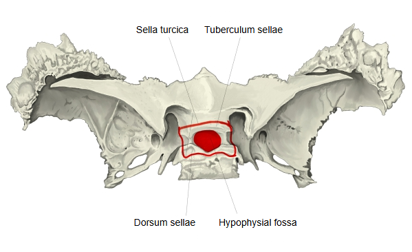
Sella turcica = tuberculum sellae (the front elevation) + hypophysial fossa (holds the pituitary gland) + dorsum sellae (the back elevation)
Some sources, however, do not include the elevations in the sella turcica; in that case
sella turcica = hypophysial fossa.
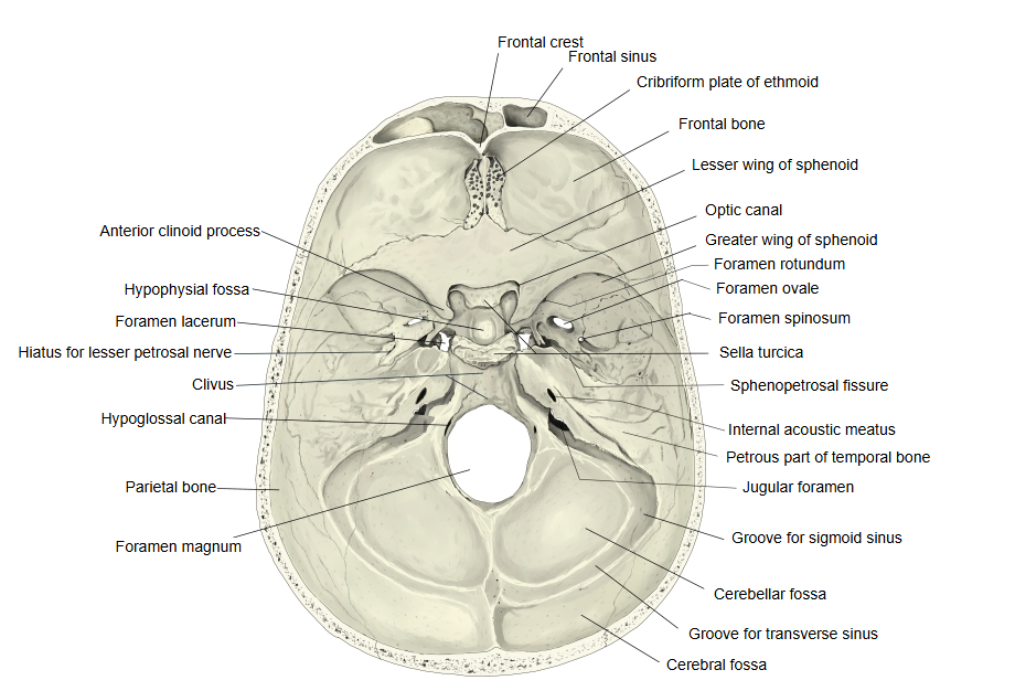
The hypophysial fossa lies in the midline of the middle cranial fossa, between the tuberculum sellae in front and the dorsum sellae behind.
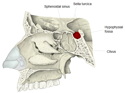
Directly below and in front of it is the sphenoidal sinus, separated from it by a thin plate of bone; behind it the bone slopes down as the clivus.
On each side lies the cavernous sinus. Above, a fold of dura mater (the diaphragma sellae) roofs it over, with a small opening for the pituitary stalk.
Because of these thin walls, the pituitary gland can be reached surgically through the nose and the sphenoidal sinus.
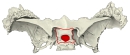 Sphenoid bone (superior view)
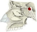 Right nasal cavity, lateral wall (section)
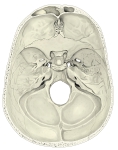 Cranial fossae (internal surface of the cranial base)
Reference websites
[Wikipedia]
· Sella turcica (hypophyseal fossa), Wikipedia.
[Websites with illustrations]
· Middle cranial fossa: boundaries and contents (TeachMeAnatomy)
· Sphenoid bone: anatomy, function and development (Kenhub)
· Sphenoid bone, upper surface (Gray's Anatomy, fig. 145) (Wikimedia Commons)

· Base of the skull, inner surface (Gray's Anatomy, fig. 193) (Wikimedia Commons)

Related terms
- Sella turcica
- Dorsum sellae
- Tuberculum sellae
- Body of sphenoid
- Sphenoidal sinus
- Pituitary gland
- Sphenoid bone
References and sources
- Further reading: Hypophyseal fossa, Wikipedia.
- Further reading: Sella turcica, Wikipedia.
- Federative International Programme for Anatomical Terminology (FIPAT). Terminologia Anatomica, 2nd ed. (TA2), 2019.