Medial supracondylar ridge (Latin: crista supraepicondylaris medialis)
Also called the medial supraepicondylar ridge or the medial supracondylar line
The name is not found in the index of some anatomy textbooks; Terminologia Anatomica calls it the medial supra-epicondylar ridge (crista supraepicondylaris medialis).
The medial border of the shaft becomes a distinct ridge in its lower third, most prominent just above the medial epicondyle. The medial intermuscular septum of the arm is attached along it and separates the front and back compartments. In front of the ridge arises brachialis and, just above the epicondyle, the humeral head of pronator teres; behind it arises the medial head of triceps brachii. It is less prominent than the lateral supracondylar ridge. Occasionally a small hooked supracondylar process projects from the front of the humerus above the medial epicondyle; a fibrous band from it can compress the median nerve or the brachial artery.
Reference websites
[Wikipedia]
· Medial supracondylar ridge, Wikipedia.
· Humerus, Wikipedia.
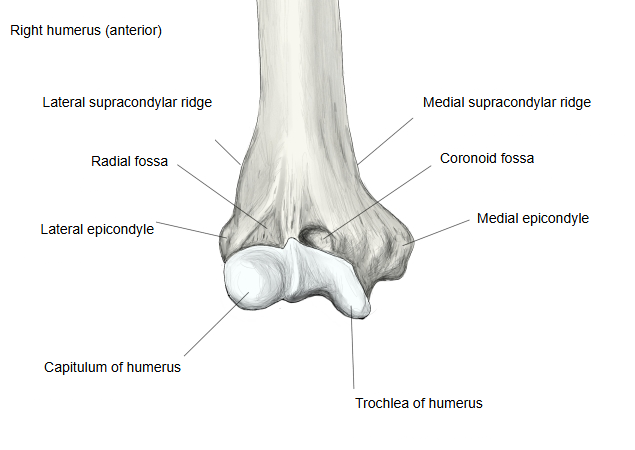
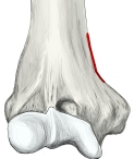 Right humerus (lower end, anterior)
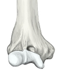 Right humerus (lower end, anterior)
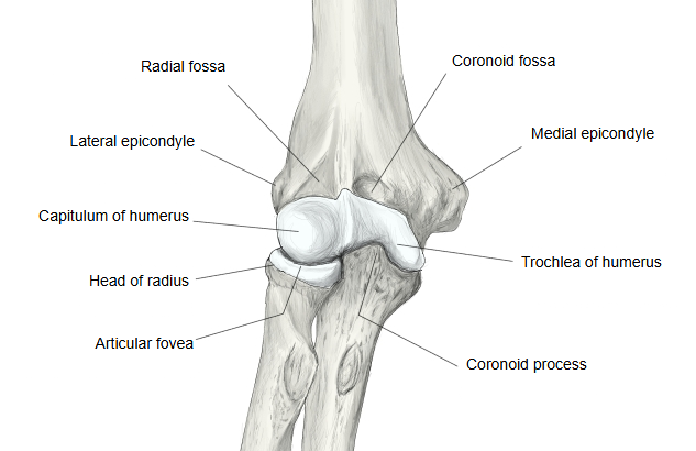
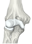 Right elbow joint (anterior)
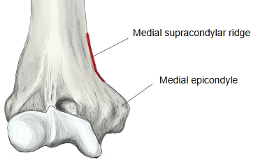
Sites with illustrations
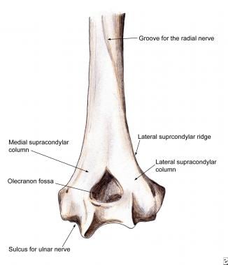
· Distal humerus, posterior view, with the medial and lateral supracondylar columns (Medscape)
Where it is found
Related terms
- Medial epicondyle
- Lateral supracondylar ridge
- Supracondylar process
- Lower end of humerus
- Groove for ulnar nerve
- Shaft of humerus
References and sources
- Further reading: Medial supracondylar ridge, Wikipedia.
- Further reading: Humerus, Wikipedia.
- Federative International Programme for Anatomical Terminology (FIPAT). Terminologia Anatomica, 2nd ed. (TA2), 2019.