Shaft of humerus (Latin: corpus humeri)
Also called the body of humerus
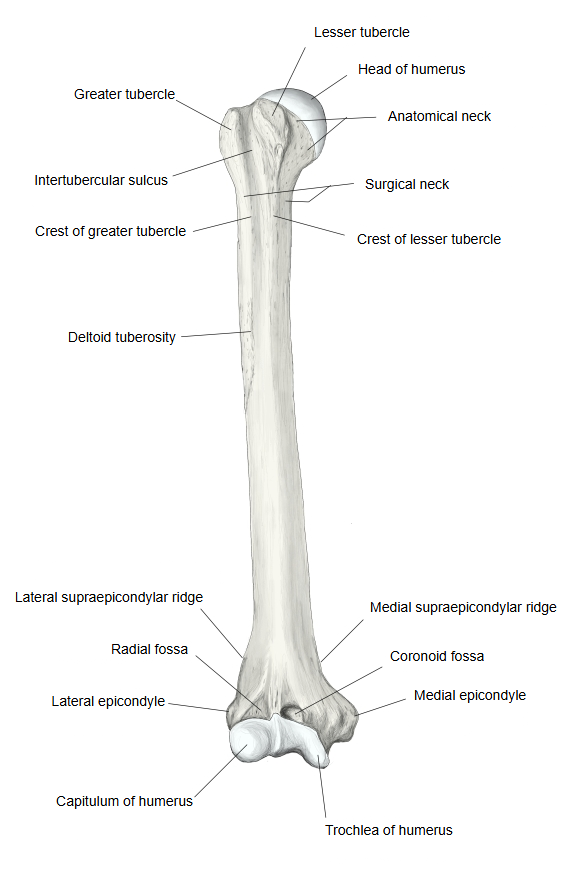
· The upper half is roughly cylindrical and the lower half triangular in section; the lowest part is flattened from front to back. It has three borders (anterior, medial and lateral) and three surfaces (anteromedial, anterolateral and posterior).
· A large nutrient foramen opens near the middle of the medial side; its nutrient canal runs downwards, towards the elbow.
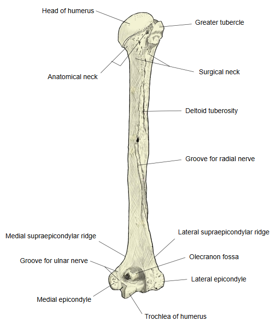
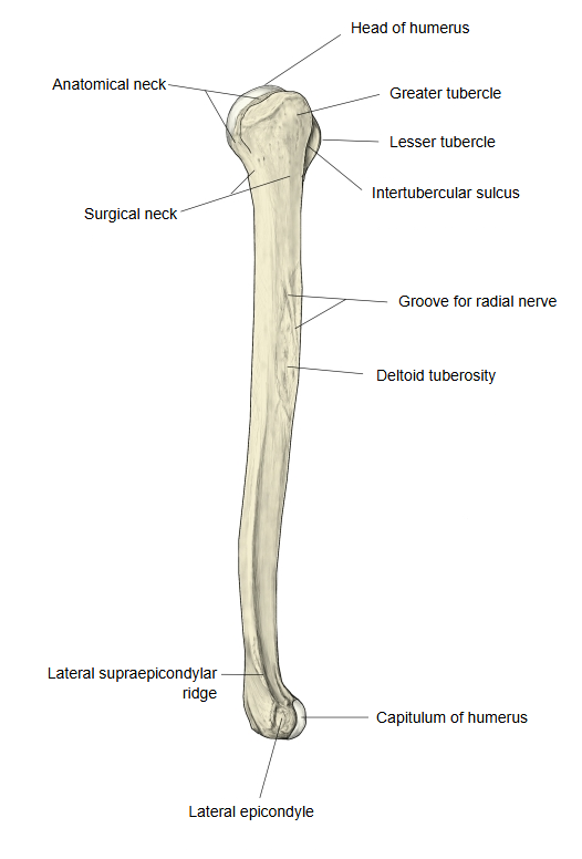
· Other named features of the shaft: the deltoid tuberosity on the lateral side near the middle, and the groove for the radial nerve, which runs obliquely downwards and laterally across the back and carries the radial nerve and the deep brachial vessels.
· Many muscles are attached to it. The main ones:
Front – origin: brachialis (lower half); insertion: deltoid, pectoralis major, teres major, coracobrachialis
Back – origin: triceps brachii (medial and lateral heads)
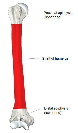
- Upper end : the expanded upper part = the proximal epiphysis.
- Shaft of the humerus : (body of the humerus) the middle part, the diaphysis of the bone.
- Lower end : the expanded lower part = the distal epiphysis.
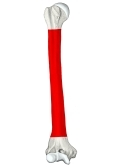 Shaft of humerus (red)
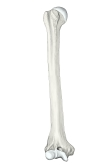 Right humerus (anterior)
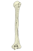 Right humerus (posterior)
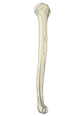 Right humerus (lateral)
Reference websites
[Wikipedia]
· Humerus, Wikipedia.
[Websites with illustrations]
· Left humerus, anterior view (Gray's Anatomy, fig. 207) (Wikimedia Commons)

· Left humerus, posterior view (Gray's Anatomy, fig. 208) (Wikimedia Commons)

· The humerus: proximal, shaft and distal (TeachMeAnatomy)
Where it is found
Related terms
- Humerus
- Proximal epiphysis of humerus
- Distal epiphysis of humerus
- Deltoid tuberosity
- Groove for radial nerve
- Surgical neck
References and sources
- Further reading: Humerus, Wikipedia.
- Further reading: Body of humerus, Wikipedia.
- Federative International Programme for Anatomical Terminology (FIPAT). Terminologia Anatomica, 2nd ed. (TA2), 2019.