Latin: plexus cervicalis
Overview
- Formed by the ventral rami of C1–C4; the dorsal rami do not take part.
- Lies deep to sternocleidomastoid, beside the transverse processes, between the prevertebral muscles and the scalenes.
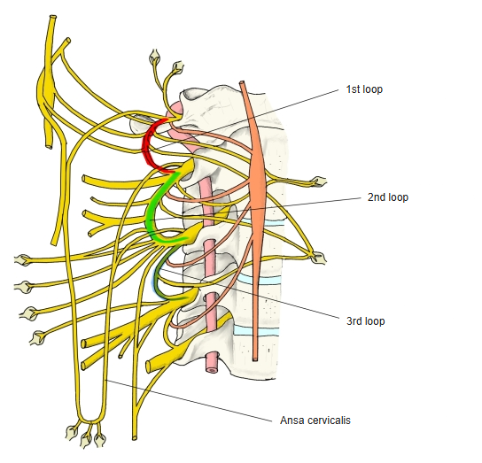
- Communicates with the accessory nerve, the hypoglossal nerve and the sympathetic trunk. The rami join in loops (first, second and third loops).
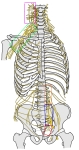
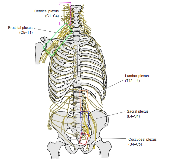
Plexuses of the spinal nerves 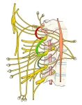
Loops of the cervical plexus
Cutaneous branches
- Four branches come out around the middle of the posterior border of sternocleidomastoid (the nerve point).
| Branch | Origin | Area of skin |
|---|---|---|
| Lesser occipital nerve | C2 (C3) | Behind the ear and the side of the back of the head |
| Great auricular nerve | C2–C3 | Over the parotid gland and the mastoid process; both surfaces of the auricle |
| Transverse cervical nerve | C2–C3 | Front of the neck |
| Supraclavicular nerves | C3–C4 | Lower neck, upper chest and shoulder |
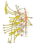
Cervical plexus (schematic)
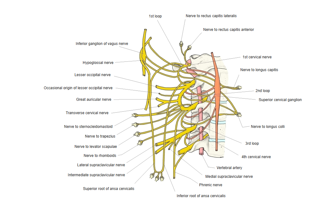
Muscular branches
| Branch | Muscles |
|---|---|
| Ansa cervicalis (C1–C3) | Sternohyoid, sternothyroid, omohyoid; C1 fibres via the hypoglossal nerve to thyrohyoid and geniohyoid |
| Phrenic nerve (C3–C5) | Diaphragm |
| Segmental muscular branches (C1–C4) | Prevertebral muscles, scalenus anterior, scalenus medius, levator scapulae |
| Branches joining the accessory nerve (C2–C4) | Sternocleidomastoid, trapezius |
References and sources
- Further reading: Cervical plexus, Wikipedia.