Latin: mamma
Overview
- A paired rounded prominence on the front of the chest, containing the mammary gland embedded in fat.
- Well developed in women after puberty; rudimentary in men.
- Base: from about the 2nd to the 6th rib, and from the sternum to the midaxillary line, mostly on the pectoralis major; an axillary tail extends toward the axilla.
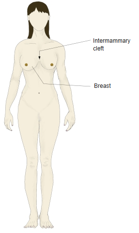
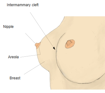
- In the centre lie the nipple and the pigmented areola; between the breasts lies the intermammary cleft.
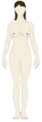
Breasts, anterior view 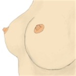
Breast, surface features
Internal structure
- The glandular tissue forms 15–20 lobes arranged radially around the nipple; each lobe is made of lobules.
- Each lobe drains by a lactiferous duct, which widens into a lactiferous sinus before opening on the nipple.
- Fat between and around the lobes gives the breast most of its size and shape.
- Suspensory ligaments (Cooper ligaments) run from the skin to the deep fascia and support the gland.
- A loose layer (retromammary space) separates the breast from the fascia of the pectoralis major.
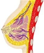
Breast, sagittal section 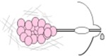
Lobe and duct system (diagram) 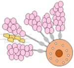
Lobes around the nipple (diagram)
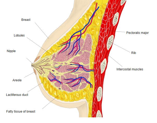
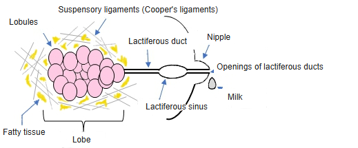
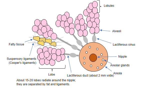
Nipple and areola
- The nipple lies at about the 4th intercostal space in men and young women; the lactiferous ducts open on its tip.
- The areola around it contains areolar glands (Montgomery glands), which form small bumps and lubricate the nipple.
- Smooth muscle in the nipple and areola makes the nipple erect.
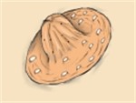
Nipple and areola
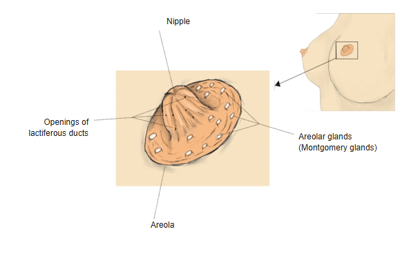
Vessels, lymph and nerves
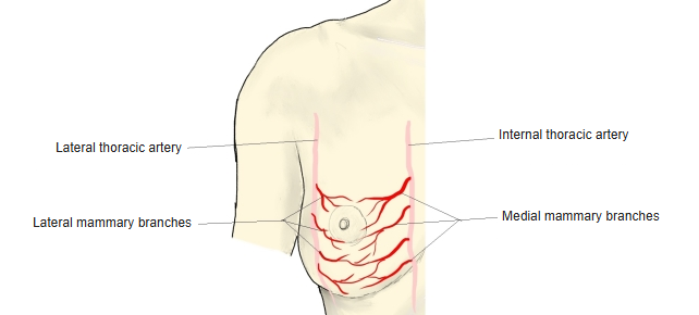
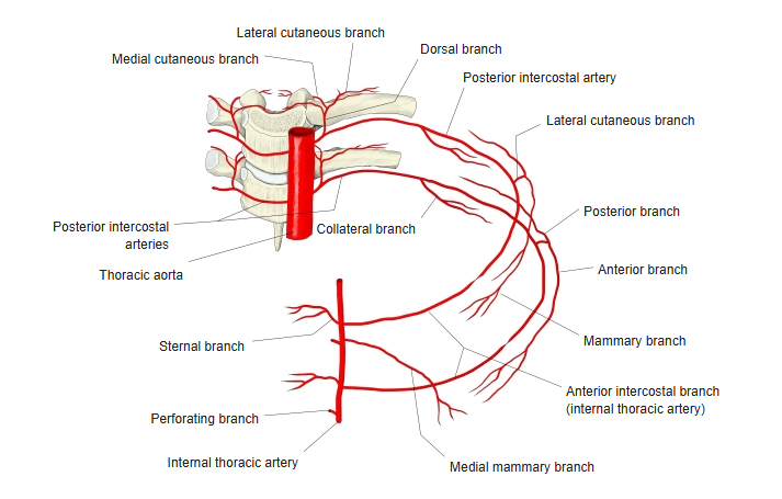
- Arteries: medial mammary branches of the internal thoracic artery, lateral mammary branches of the lateral thoracic artery, and branches of the posterior intercostal arteries.
- Veins: follow the arteries to the axillary and internal thoracic veins.
- Lymph: about 75% drains to the axillary lymph nodes; most of the rest to the parasternal nodes – important in the spread of breast cancer.
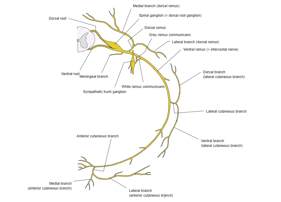
- Nerves: anterior and lateral cutaneous branches of the 4th–6th intercostal nerves.
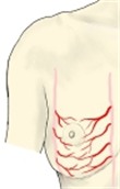
Arteries of the breast 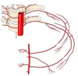
Posterior intercostal arteries 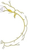
Intercostal nerve and lateral cutaneous branch
References and sources
- Further reading: Breast, Wikipedia.