Latin: palpebrae
Overview
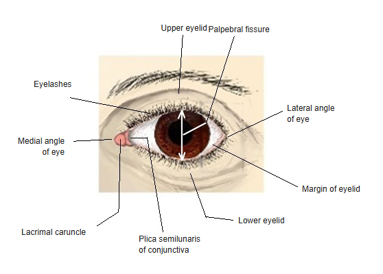
- Two movable folds, the upper and lower eyelids, that cover and protect the front of the eyeball.
- Blinking spreads tears over the cornea and pushes them towards the medial angle of the eye.
- The gap between them is the palpebral fissure; they meet at the medial and lateral angles of the eye.
- At the medial angle lie the small lacrimal lake, the reddish lacrimal caruncle and the plica semilunaris of the conjunctiva.
- A vertical skin fold that covers the medial angle (epicanthic fold) is common in some populations.
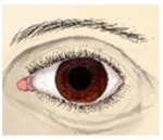
Eye, anterior view 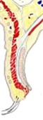
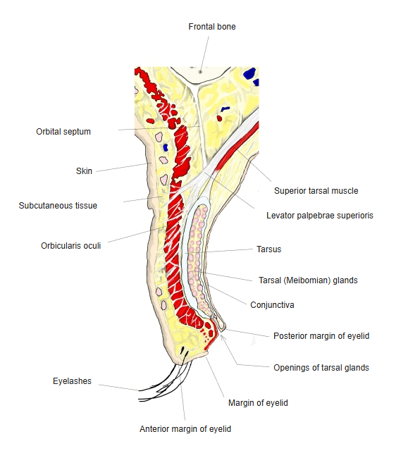
Upper eyelid, sagittal section
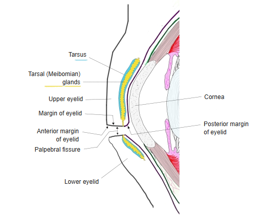
Margins of the eyelid
- Anterior margin: rounded; carries two or three rows of eyelashes, with sebaceous and modified sweat glands at their roots.
- Posterior margin: sharp, against the eyeball; a row of openings of the tarsal (Meibomian) glands lies just in front of it.
- Near the medial angle each margin has a small elevation with the lacrimal punctum, the opening of the lacrimal canaliculus.
Eyelids and tarsal glands (diagram)
Layers
| Layer (front to back) | Features |
|---|---|
| Skin | Very thin, with fine hairs and many pigment cells |
| Subcutaneous tissue | Loose, with almost no fat – it swells easily |
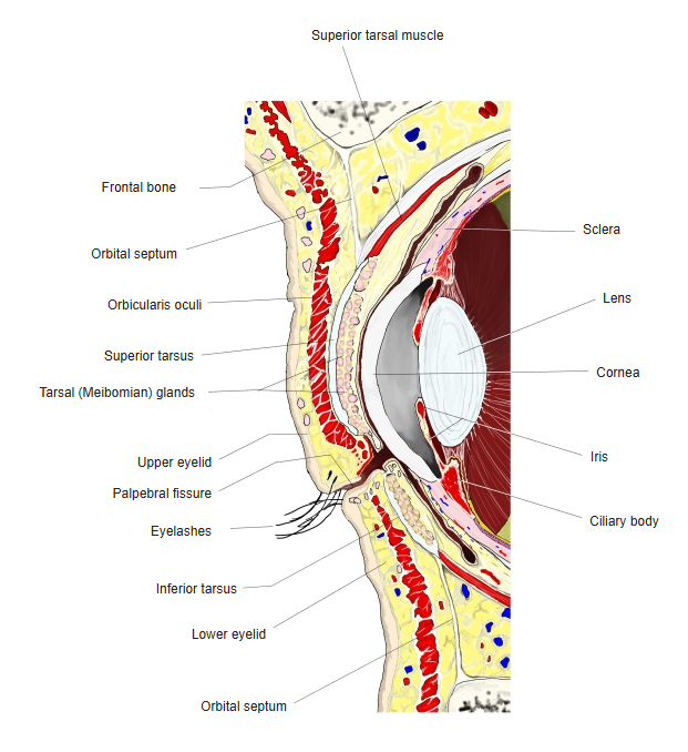
| Orbicularis oculi (palpebral part) | Thin muscle fibres that close the eyelids gently, as in blinking |
| Orbital septum | Fibrous sheet from the orbital margin to the tarsus; keeps the orbital fat in place |
| Tarsus | Plate of dense connective tissue that stiffens the lid and contains the tarsal glands, whose oily secretion slows tear evaporation |
| Conjunctiva | Palpebral conjunctiva lining the inner surface |
- In the upper eyelid the aponeurosis of the levator palpebrae superioris and the smooth superior tarsal muscle attach to the tarsus.
Eyelids, sagittal section
Vessels and nerves
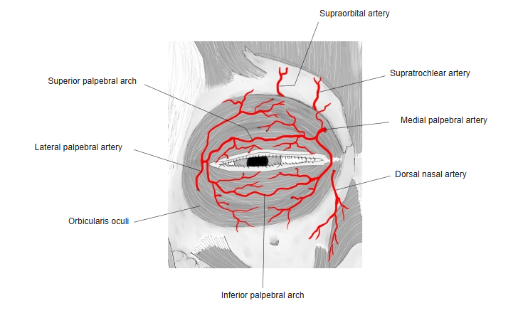
- Arteries: the medial palpebral arteries (from the ophthalmic artery) and the lateral palpebral arteries (from the lacrimal artery) join to form the superior and inferior palpebral arches.
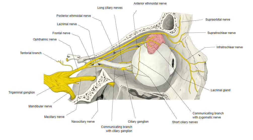
- Sensory nerves of the upper eyelid: supraorbital, supratrochlear, infratrochlear and lacrimal nerves (ophthalmic nerve).
- Sensory nerves of the lower eyelid: mainly the infraorbital nerve (maxillary nerve), medially the infratrochlear nerve.
- Motor nerves: facial nerve for orbicularis oculi; oculomotor nerve for the levator; sympathetic fibres for the superior tarsal muscle.
Arteries of the eyelids 
Nerves of the orbit
References and sources
- Further reading: Eyelids, Wikipedia.