Latin: Valva aortae
Overview
- The semilunar valve at the outflow of the left ventricle, at the root of the ascending aorta.
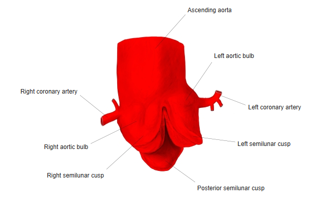
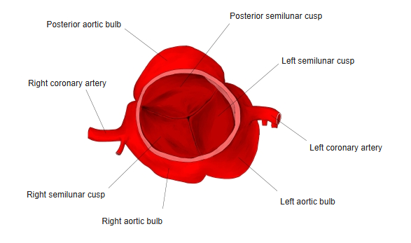
- Has three semilunar cusps: right, left and posterior (non-coronary).
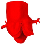
Aortic valve and aortic bulb (opened from the front) 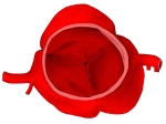
Aortic valve seen from above
Structure and function
- Above each cusp the aortic wall bulges to form an aortic sinus (aortic bulb).
- The right and left coronary arteries arise from the right and left aortic sinuses.
- Closes at the end of ventricular systole (part of the second heart sound).
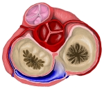
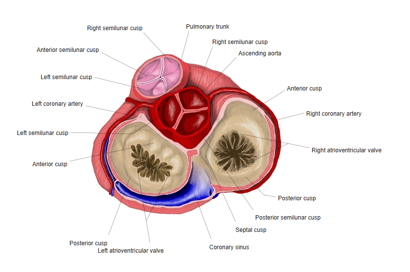
Base of the heart: the four valves seen from above
Clinical notes
- Heard best in the right 2nd intercostal space.
- Aortic stenosis (often from calcification in older people, or a bicuspid valve) and aortic regurgitation are common valve disorders.
References and sources
- Further reading: Aortic valve, Wikipedia.