Latin: Vena cava inferior
Overview
- The largest vein of the body; it returns blood from the lower limbs, pelvis and abdomen to the heart.
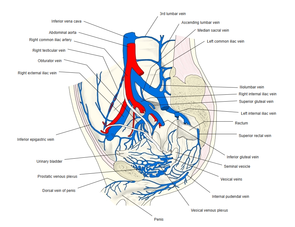
- Formed by the union of the two common iliac veins in front of the 5th lumbar vertebra, slightly to the right of the midline.
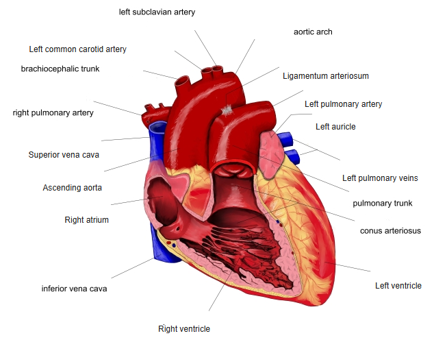
- Ascends on the right side of the abdominal aorta, passes through the caval opening of the diaphragm (T8) and ends in the lower part of the right atrium.
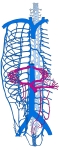
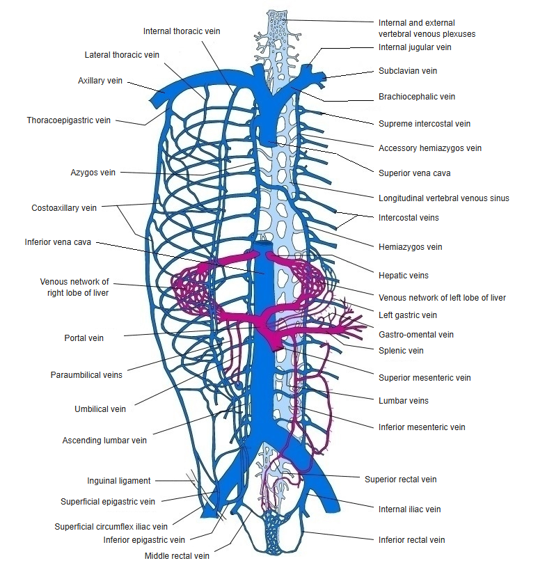
Main veins of the trunk
Course
- Runs up the posterior abdominal wall on the right of the vertebral bodies and the abdominal aorta.
- Lies in a groove on the bare area of the liver, then pierces the central tendon of the diaphragm.
- Has only a very short course in the thorax before entering the right atrium; its opening has a rudimentary valve.
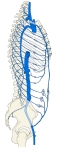
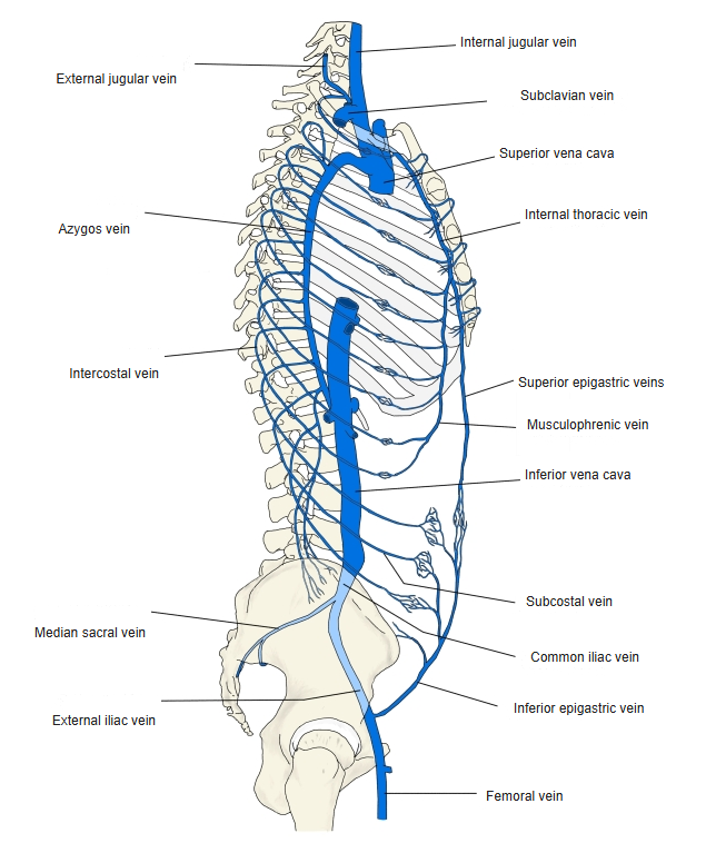
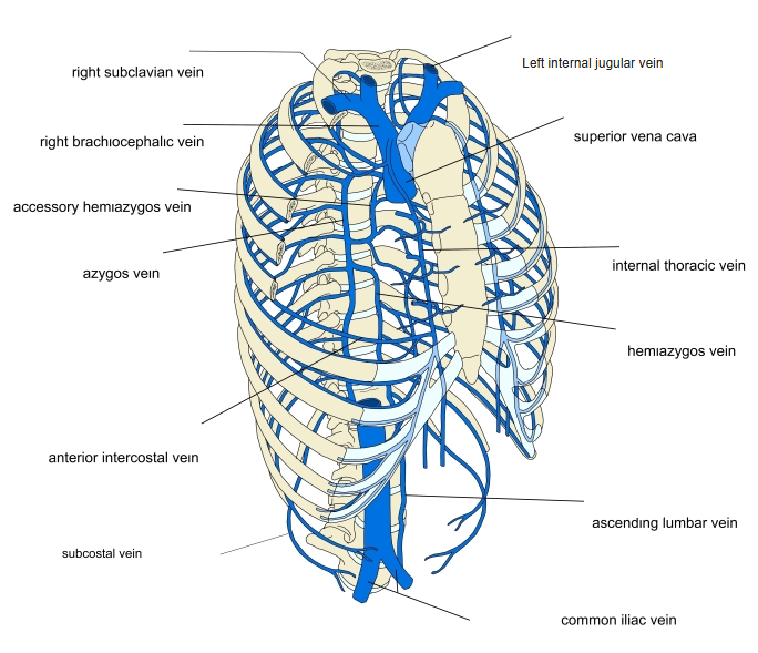
Veins of the trunk (side view) 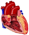
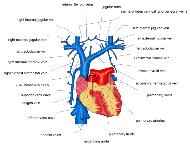
Heart and great vessels (front view) 
Veins of the male pelvis
Tributaries
| Level (approx.) | Tributaries |
|---|---|
| L5 | Common iliac veins (formation); median sacral vein |
| L1–L4 | Lumbar veins (3rd and 4th pairs); linked by the ascending lumbar veins |
| L2 | Right testicular or ovarian vein (the left one drains into the left renal vein) |
| L1 | Renal veins |
| T12–L1 | Right suprarenal vein (the left one drains into the left renal vein); inferior phrenic veins |
| T8–T10 | Hepatic veins, just below the diaphragm |
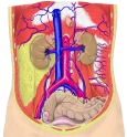
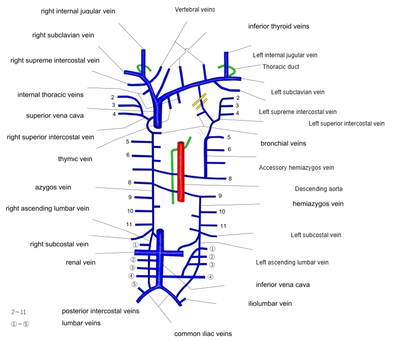
Veins of the posterior abdominal wall
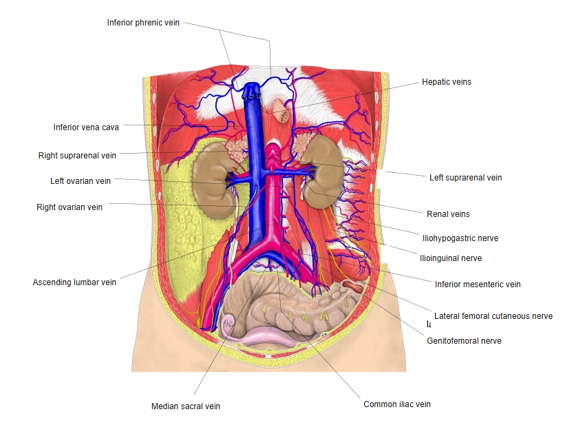
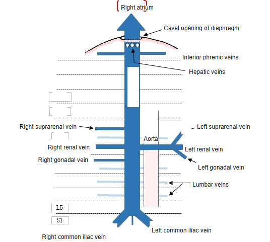
Collateral routes
- The azygos and hemiazygos veins (through the ascending lumbar veins) and the vertebral venous plexuses connect the territories of the two venae cavae.
- These routes can carry blood back to the heart when the inferior vena cava is blocked.
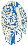
Superior vena cava and the azygos system 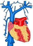
Veins entering the superior vena cava
Clinical notes
- Compression by the pregnant uterus when lying on the back can lower blood pressure (supine hypotension).
- A filter can be placed in the inferior vena cava to stop clots from the leg veins reaching the lungs.
References and sources
- Further reading: Inferior vena cava, Wikipedia.