Choroid plexus (Latin: plexus choroideus)
What is the choroid plexus?
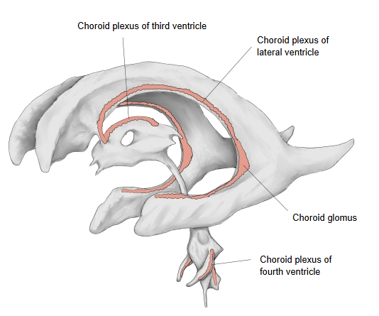
By position it can be divided into the following three:
- Choroid plexus of lateral ventricle : begins at the front end of the inferior horn and runs along the atrium and the body to the interventricular foramen, where it passes into the choroid plexus of the third ventricle. There is none in the anterior and posterior horns. Its large tuft at the atrium is the choroid glomus.
- Choroid plexus of third ventricle : two narrow strips hanging from the roof of the third ventricle; they are attached along the taenia of the thalamus (where the tela choroidea joins the thalami) and run back from the interventricular foramen.
- Choroid plexus of fourth ventricle : a right and left pair in the lower part of the roof of the fourth ventricle. They begin at the lower end of the fourth ventricle, reach the inferior medullary velum and run out into the lateral recesses, partly protruding through the lateral apertures.
Where the pia mater with its vessels (the tela choroidea) lies directly against the single layer of ependymal cells lining a ventricle, many blood vessels enter it and push folds into the ventricle, which look as if covered with small grains: this is the choroid plexus. It forms on the inner wall of the lateral ventricles, in the roof of the third ventricle and in the lower part of the roof of the fourth ventricle, behind the medulla oblongata and below the cerebellum. Its surface is a single layer of cuboidal choroid epithelium (thickened, gland-like ependymal cells) over a core of fenestrated capillaries and loose connective tissue; this epithelium secretes the cerebrospinal fluid. Tight junctions between the epithelial cells form the blood–CSF barrier.
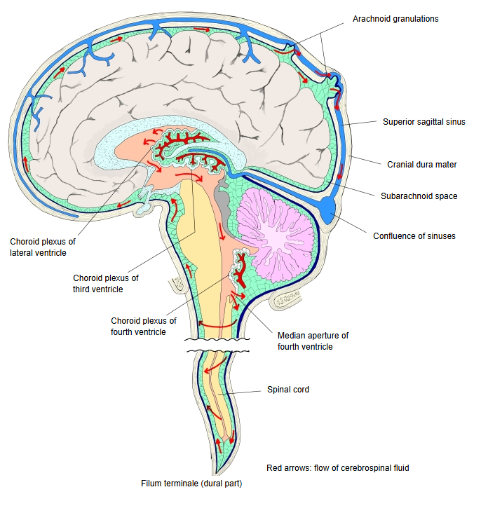
· Cerebrospinal fluid made here flows from the lateral ventricles through the interventricular foramina into the third ventricle, the cerebral aqueduct and the fourth ventricle, leaves through the median and lateral apertures into the subarachnoid space, and is absorbed mainly through the arachnoid granulations into the dural venous sinuses. Sources differ on the amounts produced each day and on the total volume; the fluid is renewed several times a day, and a smaller part of it is formed outside the choroid plexus.
Blood supply and drainage
· Lateral and third ventricles: the anterior choroidal artery (from the internal carotid artery) and the posterior choroidal arteries (from the posterior cerebral artery).
· Fourth ventricle: branches of the posterior inferior cerebellar artery.
· Veins: the superior choroid vein and the inferior choroid vein, which drain into the deep cerebral veins.
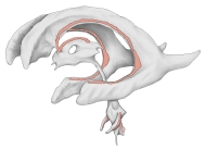 Choroid plexus in the ventricles
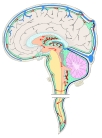 Flow of cerebrospinal fluid (median section)
Reference websites
[Wikipedia]
· Choroid plexus, Wikipedia.
· Cerebrospinal fluid, Wikipedia.
[Websites with illustrations]
· Ventricles of the brain with the choroid plexus, side and top views (diagram) (Clinical Gate)
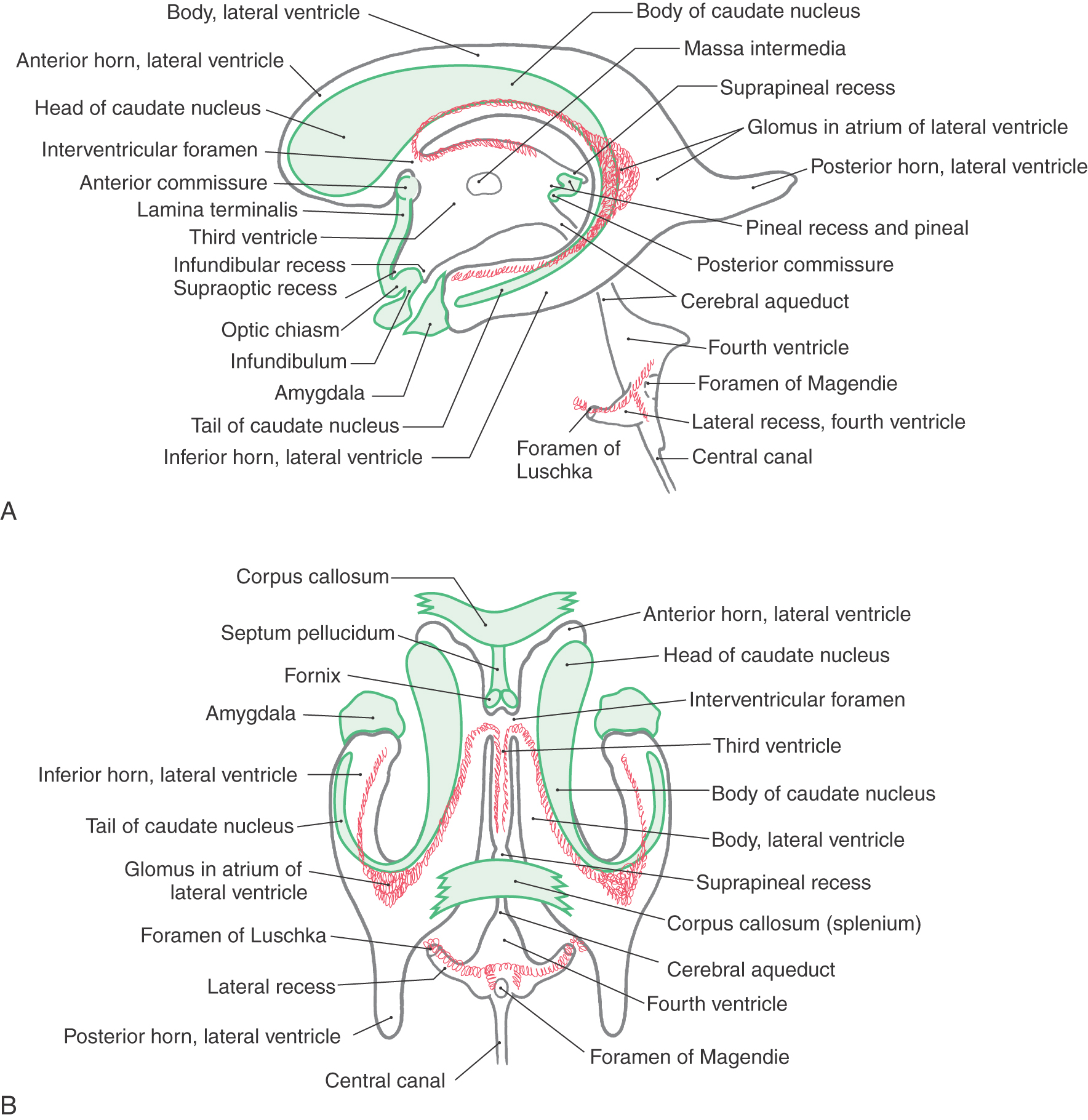
Where it is found
Related terms
- Superior choroid vein
- Inferior choroid vein
- Plexus of choroid artery
- Vein of lateral recess of fourth ventricle
- Lateral vein of lateral ventricle
References and sources
- Further reading: Choroid plexus, Wikipedia.
- Further reading: Cerebrospinal fluid, Wikipedia.
- Federative International Programme for Anatomical Terminology (FIPAT). Terminologia Anatomica, 2nd ed. (TA2), 2019.