Ependymal cell (Latin: ependymocytus)
Also called the ependymocyte
(glial cells)
- Central nervous system
- Ependymal cell : lines the ventricles, the choroid plexus and the central canal; takes part in the production and flow of cerebrospinal fluid.
- Astrocyte : star-shaped, with many processes; the most numerous glial cell of the central nervous system.
- Oligodendrocyte : one cell wraps its processes around several axons and forms their myelin sheaths.
- Microglia : small cells that act as phagocytes.
- Peripheral nervous system
- Schwann cell : around myelinated fibres it forms the myelin sheath; around unmyelinated fibres it only covers the axons. In myelinated fibres one cell serves one axon.
- Satellite cell : surrounds the nerve cell bodies in the sensory and autonomic ganglia (= capsule cell).
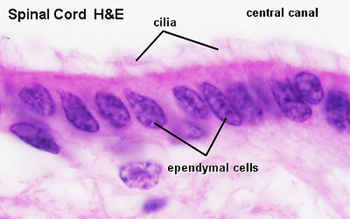
Ependymal cells are a kind of neuroglial cell that cover the inner surface of the ventricular system and the central canal of the central nervous system (brain and spinal cord). In form they resemble epithelial cells, but they differ in origin (they come from the neural tube) and function and are classed with the nervous tissue.
| Main site | Description |
|---|---|
| Inner surface of the ventricles (lateral, third and fourth ventricles) | The ependymal cells cover it as a single layer of cells. |
| Boundary between the ventricles and the central canal of the spinal cord | Take part in the movement and circulation of the cerebrospinal fluid (CSF). |
| Choroid plexus | Specialized ependymal cells produce the cerebrospinal fluid. |
| Feature | Description |
|---|---|
| Form | A single layer of epithelium-like cells (cuboidal to columnar). |
| Upper surface (towards the ventricle) | Carries cilia and microvilli; the beating cilia stir and move the CSF. |
| Basal side | Faces the underlying tissue; a complete basement membrane is often not clear. |
| Junctions | Joined mainly by adherens and gap junctions and form no tight seal, so fluid can pass between the ventricles and the brain tissue; the barrier function is limited (the ependyma is not part of the blood–brain barrier). |
| Function | Details |
|---|---|
| 1. Movement of the CSF | The beating of the cilia keeps the CSF in the ventricles circulating. |
| 2. Secretion of CSF (in the choroid plexus) | The specialized ependymal cells of the choroid plexus epithelium filter components from the capillary blood and secrete the CSF; these cells are sealed by tight junctions. |
| 3. Neural stem cell properties | Some ependymal cells may be able to give rise to new neurons or glial cells; how far this happens in the adult human brain is still debated. |
| 4. Lining of the central canal | They form the lining of the central canal of the spinal cord. |
| Type | Features |
|---|---|
| Ordinary ependymal cells | Line the ventricles and the central canal; stir the CSF with their cilia. |
| Choroid plexus epithelial cells (choroid epithelial cells) | Form the surface of the choroid plexus and produce the CSF. |
| Tanycytes | In the floor of the third ventricle; long processes link the ventricle with the hypothalamus; take part in hormone secretion and the control of metabolism. |
Clinical significance
Hydrocephalus: when the flow of CSF is blocked, for example by disorders of the ependyma or the choroid plexus, the ventricles enlarge (and the pressure in the skull rises).
Choroid plexus papilloma and ependymoma: tumours arising from the choroid plexus epithelium and from ependymal cells.
CSF shunt: an operation that drains excess cerebrospinal fluid when its flow or absorption is disturbed.
Reference websites
[Wikipedia]
· Ependyma, Wikipedia.
[Websites with illustrations]
· Ependymal cells (figure) (Biology Dictionary)
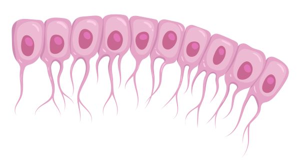
· Ependymal cells (journal figure) (Journal of Neuroinflammation (Springer Nature))

· Ependymal cells (figure) (Loony Labs)

· Spinal cord histology with the central canal (micrograph) (UNSW Embryology)
Related terms
References and sources
- Further reading: Ependyma, Wikipedia.
- Federative International Programme for Anatomical Terminology (FIPAT). Terminologia Anatomica, 2nd ed. (TA2), 2019.