Parotid papilla (Latin: papilla ductus parotidei)
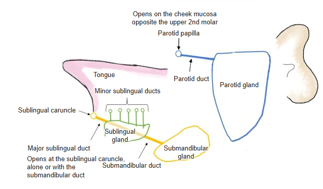
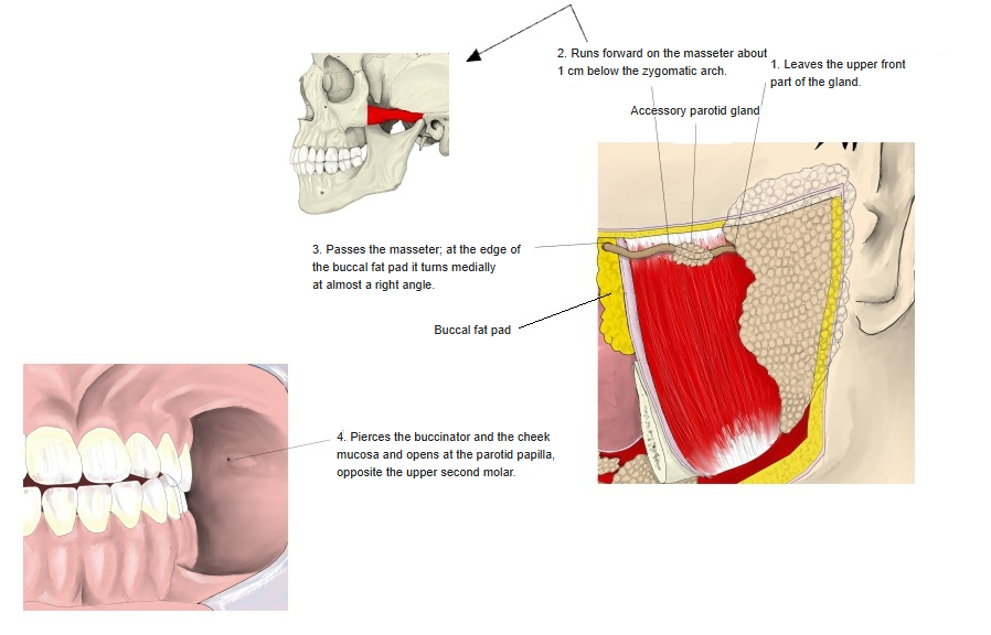
The duct of the parotid gland is the parotid duct (Stensen's duct). It leaves the front upper part of the gland and runs forwards across the outer surface of the masseter, about a fingerbreadth below and parallel to the zygomatic arch. At the edge of the buccal fat pad (the front edge of the masseter) it turns sharply inwards, pierces the buccinator and the cheek mucosa, and opens into the oral vestibule at the parotid papilla, opposite the upper second molar. An accessory parotid gland of variable size and shape is often found above the duct on the masseter; its duct joins the parotid duct. The parotid gland is pierced by the parotid plexus of the facial nerve, and also by the superficial temporal artery and the retromandibular vein. The papilla can be seen and felt in the mouth; it is the entry point for probing or imaging the duct (sialography, sialendoscopy) and may look red and swollen in mumps.
Reference websites
[Wikipedia]
· Parotid duct, Wikipedia.
· Parotid gland, Wikipedia.
[Websites with illustrations]
· Parotid papilla with the opening of the parotid duct (illustration) (Springer)

· Left parotid duct papilla (photograph) (headandneckcancercheck.com)

· Parotid papilla on the cheek mucosa (photograph) (Springer)

Where it is found
Related terms
- Parotid gland
- Accessory parotid gland
- Salivary glands
- Buccinator
- Masseter
- Zygomatic arch
- Sublingual caruncle
References and sources
- Further reading: Parotid duct, Wikipedia.
- Further reading: Parotid gland, Wikipedia.
- Federative International Programme for Anatomical Terminology (FIPAT). Terminologia Anatomica, 2nd ed. (TA2), 2019.