Head of pancreas (Latin: caput pancreatis)
· The lower left part of the head projects to the left as the uncinate process, which hooks behind the superior mesenteric vein and superior mesenteric artery. The notch where these vessels pass is the pancreatic notch.
· It lies to the right of the superior mesenteric vessels, at about the level of the second lumbar vertebra (sources give L1 to L3), and is the thickest part of the gland.
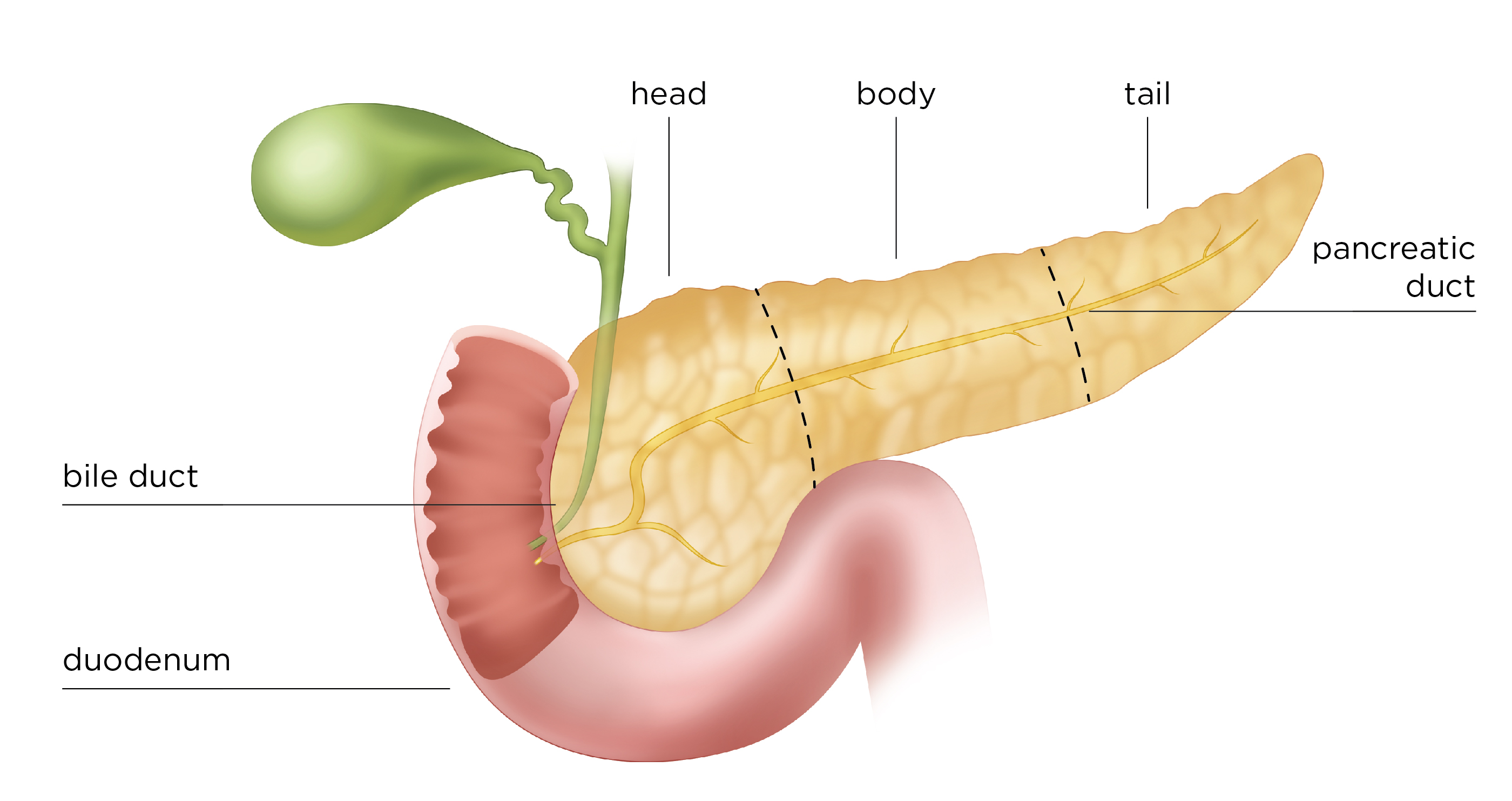
· The common bile duct runs down in a groove on, or a tunnel through, its back surface and joins the main pancreatic duct; together they open on the major duodenal papilla.
· It shares its blood supply with the duodenum through the pancreaticoduodenal arteries, which is why the two are removed together in a pancreaticoduodenectomy (Whipple operation). A tumour in the head can press on the common bile duct and cause jaundice.
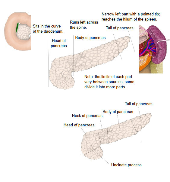
pancreas
- Head : the swollen right end, fitted into the C-shaped curve of the duodenum. Term: uncinate process (the part projecting below).
- Body : runs to the left across the front of the vertebral column; three surfaces and three borders are distinguished.
- Anterior surface : faces forward and upward, toward the back of the stomach across the omental bursa. Covered by parietal peritoneum.
- Posterior surface : flat; attached directly to the abdominal wall by loose connective tissue. Not covered by parietal peritoneum.
- Inferior surface : very narrow; faces forward and downward. Sometimes absent. Covered by parietal peritoneum.
- Anterior, superior and inferior borders : the anterior border is slightly sharp and gives attachment to the transverse mesocolon; the superior and inferior borders are blunt.
- Tail : the narrow part reaching farthest to the left; its tip is pointed and reaches about the middle of the visceral surface of the spleen, at its hilum.
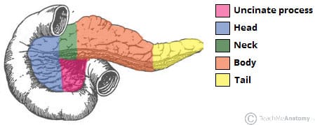
Note: some sources divide the pancreas into five parts, adding two more to the three above: the neck (a short narrow part joining the head to the body, in front of the portal vein) and the uncinate process. Most texts use four parts (head, neck, body and tail). The parts merge into one another with no sharp boundaries.
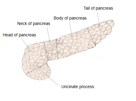
[Relations]
· In front: the transverse colon and its mesocolon, and coils of small intestine; the gastroduodenal artery runs near its upper front surface.
· Behind: the inferior vena cava, the right and left renal veins and the common bile duct.
· Around it: the first to third parts of the duodenum curve around its right side, top and bottom.
[Differences in function between the parts]
| Part | Main features and tendencies (tendencies only: there are no clear boundaries or strict limits) | |
|---|---|---|
| 1 | Head | · Especially rich in acini (exocrine tissue), important for making pancreatic juice · Close to the outlet of the main pancreatic duct (the duodenal papilla), so closely related to the bile ducts |
| 2 | Body | Both exocrine and endocrine tissue in balance |
| 3 | Tail | The islets of Langerhans are somewhat denser here, so the tail is important for the endocrine function (especially insulin secretion) |
Reference websites
[Wikipedia]
· Pancreas, Wikipedia.
· Uncinate process of pancreas, Wikipedia.
[Websites with illustrations]
· Parts of the pancreas (figure) (TeachMeAnatomy)
· Head, body and tail of the pancreas (figure) (National Pancreas Foundation)
· The pancreas (TeachMeAnatomy)
Where it is found
Related terms
- Pancreas
- Neck of pancreas
- Body of pancreas
- Tail of pancreas
- Pancreatic notch
- Duodenum
- Common bile duct
References and sources
- Further reading: Pancreas, Wikipedia.
- Further reading: Uncinate process of pancreas, Wikipedia.
- Federative International Programme for Anatomical Terminology (FIPAT). Terminologia Anatomica, 2nd ed. (TA2), 2019.