Transverse mesocolon (Latin: mesocolon transversum)
- Overview
- Structures passing through it
- Reference websites
- (Wikipedia, websites with illustrations)
- Where it is found
- Related terms
Overview
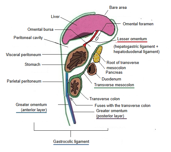
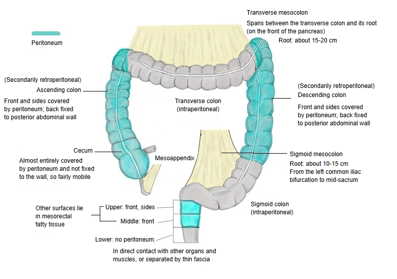
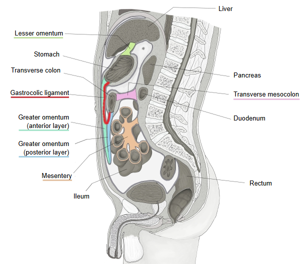
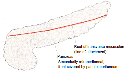
· Its root runs roughly horizontally across the front of the head and body of the pancreas. The transverse mesocolon lets the transverse colon hang freely and move.
· It divides the abdominal cavity into a supracolic compartment (liver, stomach, spleen) and an infracolic compartment (most of the small intestine). Its front fuses with the posterior layer of the greater omentum, and together they form the lower wall of the omental bursa (lesser sac).
Its size varies a great deal between individuals; rough figures are:
Width: upper border, at the root of the transverse mesocolon on the pancreas, about 15–20 cm.
Lower border, where it is attached to the transverse colon: about 30–50 cm.
Depth (from root to colon): only a few centimetres at the two ends (near the colic flexures), about 10–15 cm in the middle.
of the
peritoneum
reaching into
the abdominal
cavity
- Mesentery : a double layer of peritoneum that joins an organ to the parietal peritoneum of the wall; the path of its vessels and nerves. Examples: the mesentery of the small intestine, the transverse mesocolon and others.
- Peritoneal folds : a single layer: ridges on the inner surface of the anterior abdominal wall raised by vessels and cords. Examples: the median, medial and lateral umbilical folds.
- Omentum : peritoneum that wraps the stomach and continues up and down from its borders. Lesser omentum: stomach to liver, a double layer. Greater omentum: hangs down like a curtain; the double layer is folded back on itself, so its central part is a fused four-layered sheet.
- Peritoneal ligaments : a double layer that joins an organ to the abdominal wall or to another organ and holds it in place. Examples: the falciform ligament, the gastrosplenic ligament.
 Abdomen and pelvis (median section)
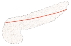 Root of transverse mesocolon
Structures passing through it
The arteries, veins, nerves and lymph nodes that run in the transverse mesocolon (mesentery) are listed below.
| Arteries | 1 | Middle colic artery | Supplies the middle part of the transverse colon with blood. It arises from the superior mesenteric artery, runs between the two layers of the transverse mesocolon to the colon and divides into right and left branches, which join the right colic and left colic arteries. |
| 2 | Right colic artery (very variable) | A branch of the superior mesenteric artery that supplies the ascending colon and the right end of the transverse colon; it joins the right branch of the middle colic artery near the right colic flexure. | |
| 3 | Left colic artery | A branch of the inferior mesenteric artery. It reaches as far as the left end of the transverse colon and joins the left branch of the middle colic artery. | |
| 4 | Marginal artery | The chain of anastomoses formed by the branches above, running along the margin of the colon; an important vessel, found close to the attached border of the transverse mesocolon. | |
| Veins | Veins accompanying the arteries, chiefly the middle colic vein, which drains into the superior mesenteric vein. | ||
| Nerves | 1 | Mainly branches of the superior mesenteric plexus | Sympathetic and parasympathetic nerve fibres running along the superior mesenteric artery pass through the transverse mesocolon with the middle colic artery. |
| 2 | Sympathetic nerve fibres | From the coeliac plexus and the superior mesenteric plexus. | |
| 3 | Parasympathetic nerve fibres | From the vagus nerve; it supplies roughly the right two-thirds of the transverse colon. The left third is supplied by the pelvic splanchnic nerves. | |
| Other | Middle colic lymph nodes | Lie within the transverse mesocolon; they collect lymph from the colon and pass it on to the superior mesenteric lymph nodes. | |
Reference websites
[Wikipedia]
· Mesentery (includes the transverse mesocolon), Wikipedia.
[Websites with illustrations]
· Transverse mesocolon (figure) (Kenhub)

· Transverse mesocolon (figure) (IMAIOS e-Anatomy)
In short, from the standard textbook description:
The base of the transverse mesocolon lies on a line running across the front of the second lumbar vertebra (some sources give the first to second); this line is the root of the transverse mesocolon. The transverse mesocolon is made of two fused layers: the posterior layer is the mesocolon proper, and the anterior layer is the continuation of the posterior layer of the greater omentum. In theory, therefore, four layers of peritoneum can be distinguished.
Where it is found
Related terms
References and sources
- Further reading: Transverse mesocolon, Wikipedia.
- Further reading: Mesocolon, Wikipedia.
- Federative International Programme for Anatomical Terminology (FIPAT). Terminologia Anatomica, 2nd ed. (TA2), 2019.