Adductor muscles (Latin: musculi adductores)
Also called the adductor group or the medial thigh muscles
the collective name for the following six muscles on the inner side of the thigh.
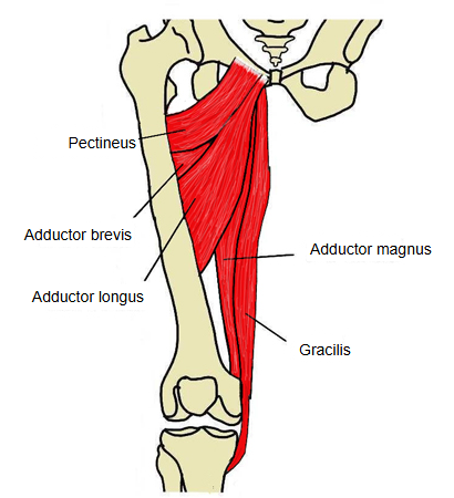
| 1 | Pectineus | the uppermost, from the pectineal line of the pubis to the pectineal line of the femur |
| 2 | Adductor brevis | behind pectineus and adductor longus, from the inferior pubic ramus to the upper linea aspera |
| 3 | Adductor longus | the most anterior, from the body of the pubis to the middle third of the linea aspera |
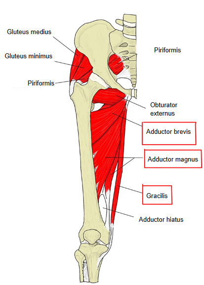
| 4 | Adductor magnus | the largest: its adductor part runs from the ischiopubic ramus to the linea aspera, its hamstring part from the ischial tuberosity to the adductor tubercle; between the two insertions is the adductor hiatus |
| 5 | Adductor minimus | the upper, more horizontal fibres of adductor magnus, often described as a separate muscle |
| 6 | Gracilis | long and strap-like, from the pubis to the medial surface of the upper tibia (part of the pes anserinus); it also flexes the knee |
They lie in the medial compartment of the thigh. They arise from the front of the pelvis (pubis and ischium, around the obturator foramen) and fan out to the femur, mostly to the linea aspera; gracilis alone reaches the tibia. Besides adduction they help to flex the hip and to stabilise the pelvis in standing and walking. Obturator externus lies deep in the same compartment and is included in the group by some sources; it acts mainly as a lateral rotator of the hip. Sources differ on the members: most English texts list pectineus, adductor longus, adductor brevis, adductor magnus and gracilis, treating adductor minimus as part of adductor magnus. Strain of the adductors at their pubic attachment (a groin strain) is a common injury; adductor longus is the one most often affected.
Most are supplied by the obturator nerve (L2–L4): adductor longus, adductor brevis, the adductor part of adductor magnus, gracilis and obturator externus. Pectineus is supplied by the femoral nerve (sometimes with a branch of the obturator or accessory obturator nerve), and the hamstring part of adductor magnus by the tibial part of the sciatic nerve. Their arteries are branches of the deep femoral artery (including its perforating branches), the obturator artery and the medial circumflex femoral artery. The femoral vessels run down between the adductors and the front of the thigh in the adductor canal.
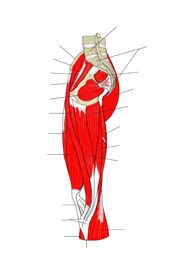 Thigh muscles (medial view)
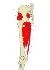 Deep adductors (anterior view)
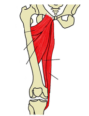 Adductor group (anterior view)
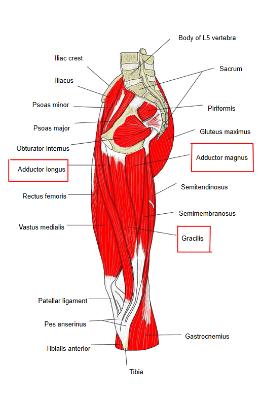
Where it is found
Related terms
References and sources
- Further reading: Adductor muscles of the hip, Wikipedia.
- Federative International Programme for Anatomical Terminology (FIPAT). Terminologia Anatomica, 2nd ed. (TA2), 2019.