Bronchiole (Latin: bronchiolus)
Also called the plural bronchioles (bronchioli) or the lobular bronchiole
1. Diameter: about 0.8–1 mm (sources differ; usually given as about 1 mm or less).
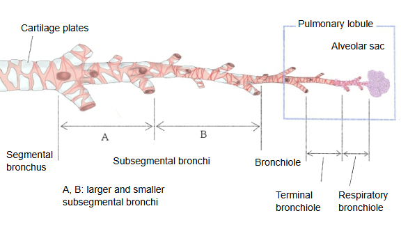
2. The cone-shaped region, about 1–2 cm across, supplied by one bronchiole is called a pulmonary lobule.
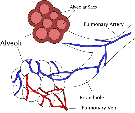
3. Inside the pulmonary lobule it divides into 3–6 branches (commonly 3–5 terminal bronchioles), which pass on to the next order of airway: respiratory bronchioles, alveolar ducts and alveolar sacs.
4. In the larger bronchioles the lining is a pseudostratified ciliated epithelium with goblet cells. Further along it becomes ciliated cuboidal epithelium: the goblet cells become few and disappear, and club cells (formerly Clara cells) appear.
5. In the largest bronchioles, older descriptions such as this one still find bronchial glands (mixed glands) in the lamina propria; most modern texts define the bronchiole as having no glands.
6. Beneath the lamina propria runs a relatively thick layer of spirally arranged smooth muscle.
7. Beneath the muscle layer is a layer of loose connective tissue, in which, in the largest bronchioles, small pieces of cartilage may still be found.
8. Once the diameter is 1 mm or less, no cartilage (bronchial cartilages) is seen, and the smooth muscle becomes well developed and runs round the airway. Without cartilage, the bronchiole is held open by elastic fibres that tie its wall to the surrounding lung tissue, so it widens as the lung expands.
The smooth muscle controls airflow: sympathetic stimulation and adrenaline widen the bronchioles (bronchodilation), while parasympathetic (vagal) stimulation, histamine and irritants narrow them (bronchoconstriction), as in asthma.
From the bronchi to the alveoli
Reference websites
[Wikipedia]
· Bronchiole, Wikipedia.
· Club cell, Wikipedia.
[Websites with illustrations]
· Bronchiole and alveoli (diagram) (Wikimedia Commons)
· Bronchioles (illustration) (Science Photo Library)

· Bronchioles and alveoli (diagram) (Medicine LibreTexts)

Where it is found
Related terms
References and sources
- Further reading: Bronchiole, Wikipedia.
- Further reading: Club cell, Wikipedia.
- Federative International Programme for Anatomical Terminology (FIPAT). Terminologia Anatomica, 2nd ed. (TA2), 2019.