Alveolar duct (Latin: ductus alveolaris)
Also called the plural alveolar ducts
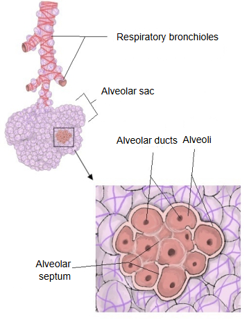
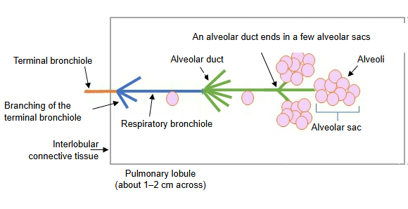
The main features of the alveolar duct are summarised below.
1. Each alveolar duct has 20–60 alveoli opening from its wall.
2. In a longitudinal section of the duct wall, the epithelium no longer forms a continuous lining: it is interrupted by the openings of the alveoli.
3. Between the openings of neighbouring alveoli, the wall of the duct remains only as small islands or knob-like rims.
4. The epithelium consists of undifferentiated cuboidal cells (probably a transitional form), but nearer the peripheral alveolar sacs the rounded and flat cells seen in the alveoli (type II and type I alveolar cells) appear.
5. Under the epithelium, spiral smooth muscle, collagen fibres and elastic fibres form a supporting layer. The elastic fibres stretch during inspiration and help the lung recoil during expiration.
6. No branch of the pulmonary artery runs with it; only capillaries accompany it.
7. At the end of each alveolar duct, 2–5 alveolar sacs form a grape-like cluster.
8. The part that connects the alveolar duct with the alveolar sacs is called the atrium. Sources differ: some describe it as a separate space between the duct and the sacs, others treat it as the end of the duct.
9. The mouth of each alveolar sac is encircled by a ring of smooth muscle, collagen fibres and elastic fibres.
From the bronchi to the alveoli
Reference websites
[Wikipedia]
· Alveolar duct, Wikipedia.
· Pulmonary alveolus, Wikipedia.
[Websites with illustrations]
· The respiratory zone (OpenStax Anatomy and Physiology)

· Alveolar duct and alveoli (diagram) (Study.com)

· Alveolar duct (picture) (Know Your Body)

Where it is found
Related terms
References and sources
- Further reading: Alveolar duct, Wikipedia.
- Further reading: Pulmonary alveolus, Wikipedia.
- Federative International Programme for Anatomical Terminology (FIPAT). Terminologia Anatomica, 2nd ed. (TA2), 2019.