Pancreatic islets (Latin: insulae pancreaticae)
Also called Islets of Langerhans
- Overview
- Questions and answers
- Additional points
- History
- Reference websites
- (Wikipedia, websites with illustrations)
- Where it is found
- Related terms
Overview
structure
of the
pancreas
- Exocrine part
- Acini : made of acinar cells; about 80–85% of the pancreas.
- Ducts : made of duct epithelial cells and centroacinar cells; about 5–10%.
- Endocrine part : the pancreatic islets (islets of Langerhans), which secrete hormones; about 1–2%.
- β (beta) cells : secrete insulin; about 60–70% of the islet cells.
- α (alpha) cells : secrete glucagon; about 15–25%.
- δ (delta) cells : secrete somatostatin; about 5–10%.
- F (PP) cells : secrete pancreatic polypeptide; about 1–5%.
- ε (epsilon) cells : secrete ghrelin; less than 1%.
- Others : connective tissue, blood vessels, nerves, stroma, immune cells and others; about 1–3%.
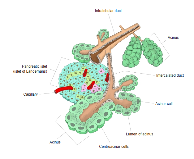
[Cell types of the islets]
| Cell | Share (approx.) | Secretes | Action | |
|---|---|---|---|---|
| 1 | β cells (beta) | 60–70% | Insulin | Lowers blood glucose (promotes the uptake of glucose into cells) |
| Size: about 10–20 µm. Shape: polygonal to slightly rounded. Other: central nucleus, many secretory granules. | ||||
| 2 | α cells (alpha) | 15–25% | Glucagon | Raises blood glucose (promotes the breakdown of glycogen in the liver) |
| Size: about the same as β cells. Shape: slightly oval. Other: more often found toward the edge of the islet than β cells. | ||||
| 3 | δ cells (delta) | 5–10% | Somatostatin | Inhibits the release of insulin and glucagon |
| Size: slightly smaller. Shape: irregular. Other: granules relatively small and dense. | ||||
| 4 | F cells (PP cells) | 1–5% | Pancreatic polypeptide | Regulates the movements of the gastrointestinal tract and the secretion of pancreatic juice |
| Size: small. Shape: irregular. Other: a few at the edge of the islet, secretory granules fine and scattered; most common in islets of the head of the pancreas. | ||||
| 5 | ε cells (epsilon) | very few | Ghrelin | An appetite-stimulating hormone |
| Size: the smallest. Shape: irregular to slightly spindle-shaped. Other: very few; more numerous before birth, very rare in adults. | ||||
Questions and answers
Q: Do the proportions of the cell types differ with the region of the pancreas?
A: Yes. The proportions of the islet cells (β, α, δ and others) differ between the tail, body and head of the pancreas. The clearest differences are in the density of the islets and of β cells, and in the PP cells.
| Part of the pancreas | β cells | α cells | Remarks |
|---|---|---|---|
| Tail | many (highest density) | relatively many | The main centre of insulin secretion; the islets are densest here. |
| Body | intermediate | intermediate | Typical composition. |
| Head | fewer | fewer (posterior part) | Islets of the posterior head and uncinate process are rich in PP cells and poor in glucagon-secreting α cells. |
Additional points
· β cells (insulin secretion) are most plentiful in the tail (left end) of the pancreas, where the islets are densest. This is an important fact in diabetes research and treatment.
· α cells (glucagon secretion) are not more numerous in the head: the islets of the posterior head and uncinate process, which develop from the ventral pancreatic bud, have few α cells, while α cells are relatively more common in the body and tail.
· δ cells are spread fairly evenly; PP cells show the clearest regional difference, being concentrated in the islets of the posterior head.
Q: What is found in the islets besides the endocrine cells?
A: The islets are clusters of endocrine cells that secrete hormones, but besides these cells they contain several important structures and components.
| Component | Outline and role | |
|---|---|---|
| 1 | Capillaries (fenestrated) | The route by which endocrine hormones (insulin, glucagon and others) are released quickly into the blood; they are in close contact with the cells. |
| 2 | Nerve endings (autonomic) | Sympathetic and parasympathetic nerve endings enter the islet and regulate the secretion of hormones by the cells. |
| 3 | Basement membrane and extracellular matrix (ECM) | Lie between the endocrine cells and the capillaries; they support the cells and influence the diffusion of hormones. |
| 4 | Connective tissue (very little) | A small amount of supporting tissue that helps to divide the structure: collagen, fibronectin and others. |
| 5 | Immune cells (a few) | Very few in a normal islet, mainly macrophages; they increase in diseases such as diabetes. |
[Clinical notes]
· Loss of β cells (type 1) or reduced insulin effect and secretion (type 2) causes diabetes mellitus.
· Tumours of islet cells (for example insulinoma) can release excess hormone.
History
The islets were first described in 1869 by the German medical student Paul Langerhans (1847–1888), then 22 years old, who found them while studying the pancreas under the microscope; hence the name islets of Langerhans. Their function was then unknown: an endocrine role was proposed only in the 1890s, and insulin was isolated in 1921–22. The pancreas contains about a million islets, each about 0.1–0.2 mm across, which together form about 1–2% of its volume. Three main cell types were described first: A (α) cells, which make glucagon and raise blood glucose; B (β) cells, which make insulin and lower it; and D (δ) cells, which make somatostatin and restrain both.
Reference websites
[Wikipedia]
· Pancreatic islets, Wikipedia.
· Pancreas, Wikipedia.
[Websites with illustrations]
· Pancreatic islet (figure) (ScienceDirect (Elsevier))

· Pancreatic islet and acini (figure) (ScienceDirect (Elsevier))

· The pancreas (TeachMeAnatomy)
Where it is found
Related terms
References and sources
- Further reading: Pancreatic islets, Wikipedia.
- Further reading: Pancreas, Wikipedia.
- Federative International Programme for Anatomical Terminology (FIPAT). Terminologia Anatomica, 2nd ed. (TA2), 2019.