Cubital fossa (Latin: fossa cubitalis)
Also called: antecubital fossa, elbow pit
The cubital fossa
It lies in front of the elbow joint, between two forearm muscles: pronator teres, which arises from the medial epicondyle of the humerus, and brachioradialis, which arises from the lateral supracondylar ridge above the lateral epicondyle. It is the usual route by which the main artery and a major nerve of the arm pass into the forearm. The superficial veins over it are the most common site for venepuncture (taking blood) and for placing intravenous lines.
Vessels and nerves passing through the cubital fossa
In the cubital fossa the brachial artery divides into the radial artery and the ulnar artery. These arteries have accompanying veins, so the brachial, radial and ulnar veins run alongside them.
Superficial veins run in the subcutaneous tissue of the cubital fossa: the cephalic vein, the basilic vein, the median cubital vein that joins them, and often the median antebrachial vein with its median cephalic and median basilic branches. These veins are used for injections and for taking blood. Their pattern varies a great deal between people, so the best place to take blood differs from person to person. The median nerve also passes through the fossa.
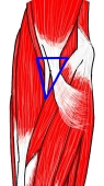 Muscles around the fossa
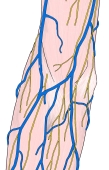 Superficial veins and nerves
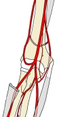 Arteries of the upper limb
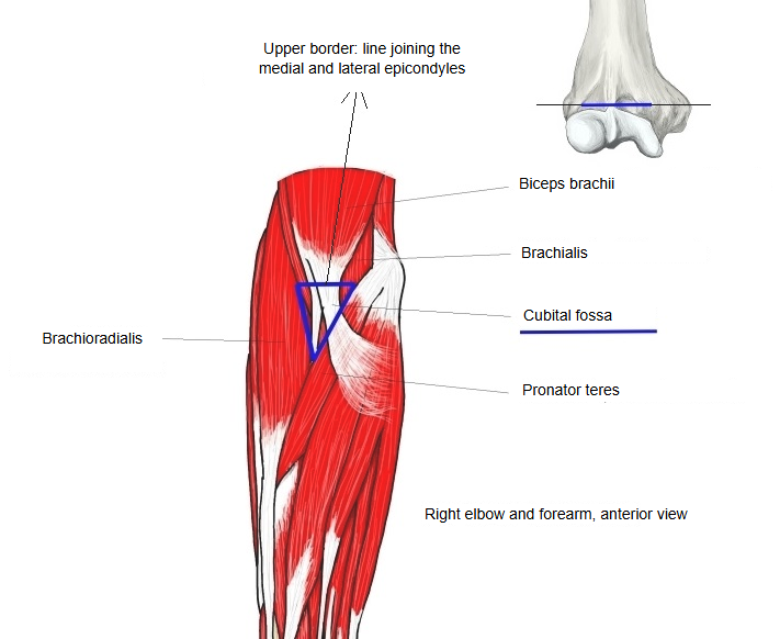
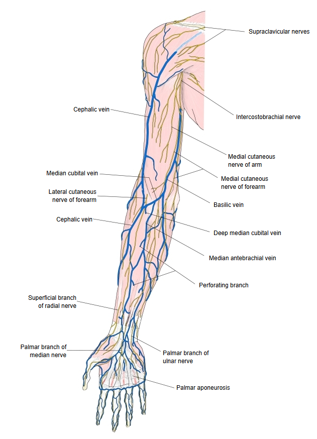
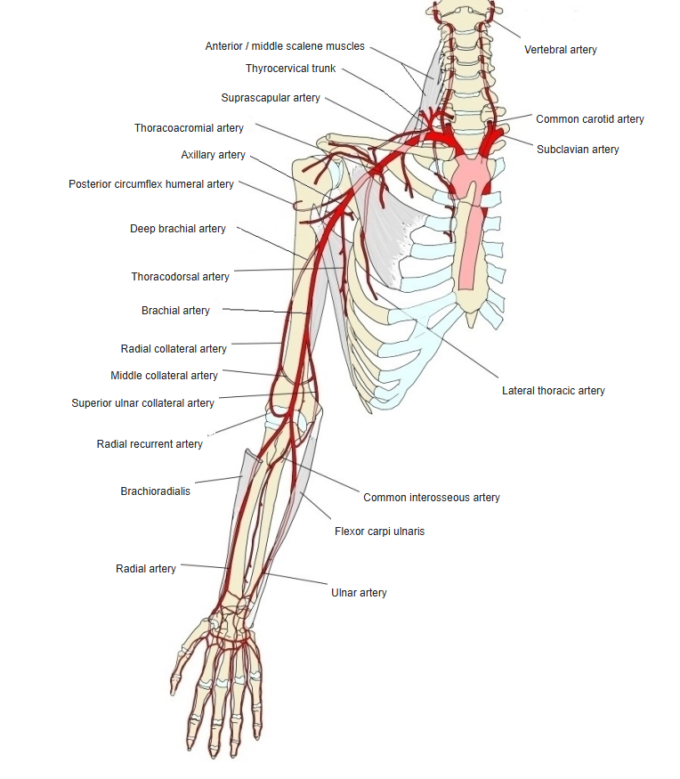
The apex of the triangle points downwards, where pronator teres and brachioradialis meet. The deep fascia of the roof is strengthened by the bicipital aponeurosis, a sheet from the tendon of biceps brachii; it lies between the median cubital vein and the brachial artery and gives the artery some protection during venepuncture.
The contents, from lateral to medial, are the radial nerve (at the lateral edge, under brachioradialis), the tendon of biceps brachii, the brachial artery and the median nerve. The brachial artery usually divides at about the level of the neck of the radius; the radial nerve divides into its superficial and deep branches near the lateral epicondyle. The brachial pulse can be felt just medial to the biceps tendon, where a stethoscope is placed when measuring blood pressure.
Reference websites
[Wikipedia]
· Cubital fossa, Wikipedia.
· Median cubital vein, Wikipedia.
[Websites with illustrations]
· The cubital fossa: borders and contents (TeachMeAnatomy)
· Cubital fossa (slide 4) (SlideShare)

· Cubital fossa (slide 10) (SlideShare)

Where it is found
Related terms
- Median cubital vein
- Brachial artery
- Median nerve
- Biceps brachii
- Pronator teres
- Brachioradialis
- Medial epicondyle
References and sources
- Further reading: Cubital fossa, Wikipedia.
- Further reading: Median cubital vein, Wikipedia.
- Federative International Programme for Anatomical Terminology (FIPAT). Terminologia Anatomica, 2nd ed. (TA2), 2019.