Diploic canals (Latin: canales diploici)
The table below shows, in simple form, the three layers of the bones of the calvaria.
| 1 | Outer table | The outermost layer, made of thick compact bone. |
| 2 | Diploe | The middle layer, made of spongy bone. It contains the diploic canals, through which veins run (diploic veins and emissary veins). |
| 3 | Inner table | The innermost layer, made of thin compact bone. On its inner surface are: 1. Digital impressions: made by the cerebral gyri 2. Cerebral juga: ridges matching the cerebral sulci 3. Arterial and venous grooves: made by the arteries and veins 4. Granular foveolae: pits that hold the arachnoid granulations of the arachnoid mater |
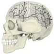 Diploe and diploic canals
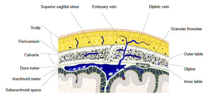 Layers of the scalp, calvaria and meninges
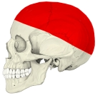 Calvaria (lateral view)
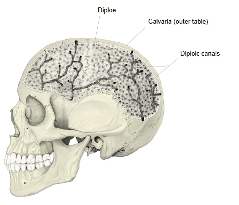
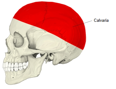
The diploic canals are channels in the diploe of the skull that hold the diploic veins. They branch through the diploe and can be seen on X-rays or when the outer table is removed. The diploic veins in them have no valves; they drain to the dural venous sinuses and, through emissary veins (for example the parietal emissary vein of the parietal bone), to veins of the scalp. The main named diploic veins are the frontal, anterior temporal, posterior temporal and occipital diploic veins.
Reference websites
· Scalp, calvaria and meninges with a diploic vein (Netter figure), ScienceDirect (Elsevier).

· Diploic and emissary veins of the skull (Netter figure), Netter Images.

Related terms
- Calvaria
- Diploic veins
- Frontal diploic vein
- Occipital diploic vein
- Parietal emissary vein
- Parietal bone
- Granular foveolae
References and sources
- Further reading: Diploë, Wikipedia.
- Further reading: Diploic veins, Wikipedia.
- Federative International Programme for Anatomical Terminology (FIPAT). Terminologia Anatomica, 2nd ed. (TA2), 2019.