Nasal mucosa (Latin: tunica mucosa nasi)
Also called the nasal mucous membrane
- Overview
- Respiratory region
- Olfactory region
- Reference websites
- (Wikipedia, websites with illustrations)
- Related terms
Overview
mucosa
- Respiratory region : the mucosa that covers most of the nasal cavity; rich in blood vessels and pale red. Part of the respiratory system.
- Olfactory region : the part at the top of the nasal cavity that contains the olfactory cells; yellowish. Part of the olfactory (sensory) system.
· It is continuous with the skin of the nasal vestibule at the front, with the lining of the paranasal sinuses, and through the choanae with the mucosa of the nasopharynx. It is firmly bound to the periosteum and perichondrium of the walls and conchae.
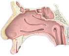 Mucosa of the lateral wall
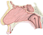 Mucosa of the nasal septum
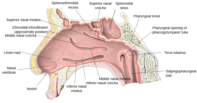
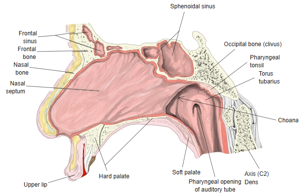
Respiratory region
The airway
Features (sources: Wikipedia and anatomy textbooks)
1. 1. It is rich in blood vessels and pinkish red.
2. 2. Over the inferior and middle nasal conchae it forms venous plexuses (the cavernous plexuses of the conchae), which can fill and empty, making the mucosa swell or shrink.
3. 3. Its lamina propria contains lymphocytes, lymphoid nodules and many nasal glands, which secrete the nasal mucus.
4. 4. The front lower part of the nasal septum is the commonest site of nosebleeds and is called Kiesselbach's area.
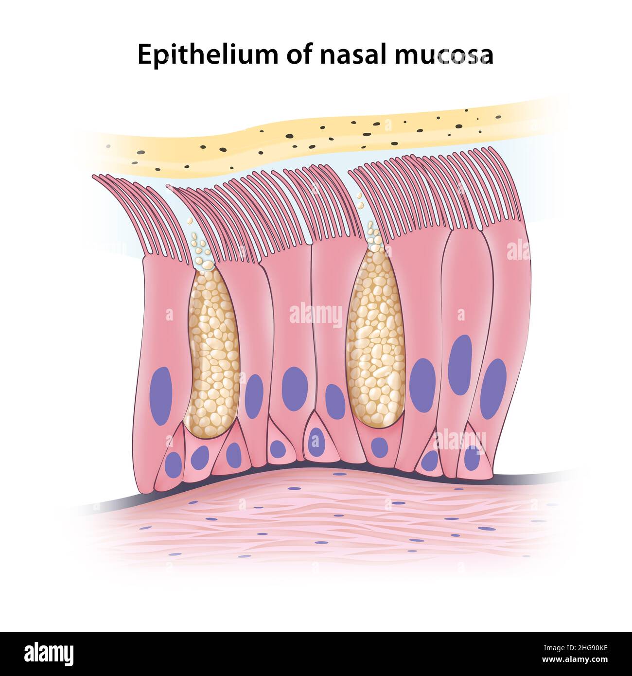
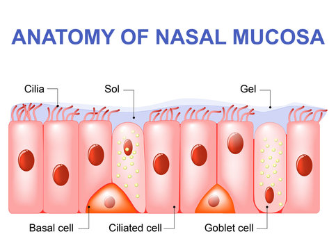
5. 5. The epithelial cells are columnar and ciliated (pseudostratified ciliated columnar epithelium); the basal cells at its base are pyramidal.
6. 6. Goblet cells (mucus-secreting cells) are scattered between the columnar cells.
· The respiratory region warms, moistens and cleans the inspired air.
· Website with an illustration of the structure of the respiratory region - 1
· Website with an illustration of the structure of the respiratory region - 2
Olfactory region
Features (sources: Wikipedia, anatomy textbooks and other websites)
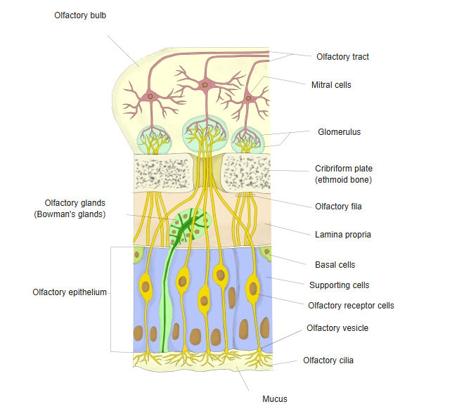
1. 1. The epithelial cells are columnar, without the motile cilia of the respiratory region; the olfactory cells carry non-motile olfactory cilia.
2. 2. The main cells of the epithelium (olfactory epithelium) are the olfactory cells and the supporting cells (with oval nuclei), with basal cells at its base.
3. 3. Small granules of yellow (yellowish-brown) pigment are seen in the surface part of the epithelium.
4. 4. The lamina propria contains the olfactory glands (Bowman's glands), whose watery secretion keeps the surface moist and dissolves odour molecules.
· Website with an illustration of the structure of the olfactory region - 1

· Website with an illustration of the structure of the olfactory region - 2

Reference websites
[Wikipedia]
· Nasal mucosa, Wikipedia.
· Olfactory epithelium, Wikipedia.
[Websites with illustrations]
· Nasal mucosa (illustration) (MedlinePlus)

· Nasal mucosa (Wikimedia Commons)

Related terms
- Nasal cavity
- Nasal glands
- Nasal septum
- Nasal vestibule
- Upper respiratory tract
- Olfactory epithelium
- Olfactory receptor cells
References and sources
- Further reading: Nasal mucosa, Wikipedia.
- Further reading: Olfactory epithelium, Wikipedia.
- Federative International Programme for Anatomical Terminology (FIPAT). Terminologia Anatomica, 2nd ed. (TA2), 2019.