Thoracic cavity (Latin: cavitas thoracis)
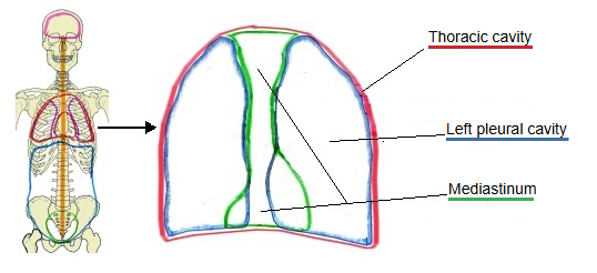
The thoracic cavity is the space enclosed by the thoracic wall and the diaphragm. It can be divided into the right and left pulmonary regions, which hold the lungs, and the mediastinum, the middle part between them.
Thoracic wall: the sternum, ribs and thoracic vertebrae with their muscles; the cavity opens upwards at the superior thoracic apertureEach lung lies in its own pleural sac
Structure
of the
thoracic
cavity
of the
thoracic
cavity
- Pulmonary regions : right and left; each holds a lung, surrounded by the pleural cavity (the space between the visceral and parietal pleura).
- Mediastinum
- Superior : thymus, brachiocephalic veins, superior vena cava (upper part), aortic arch, trachea and others.
- Inferior : heart, superior vena cava (lower part) and the end of the inferior vena cava, pulmonary veins, main bronchi and others.
- Common to both : oesophagus and vagus nerves; sympathetic trunks and aorta; azygos vein and thoracic duct.
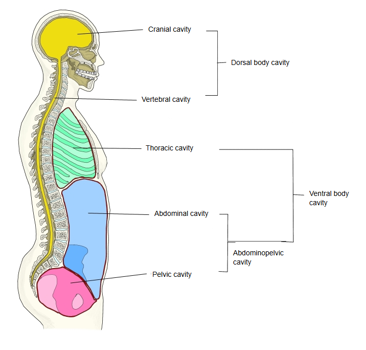
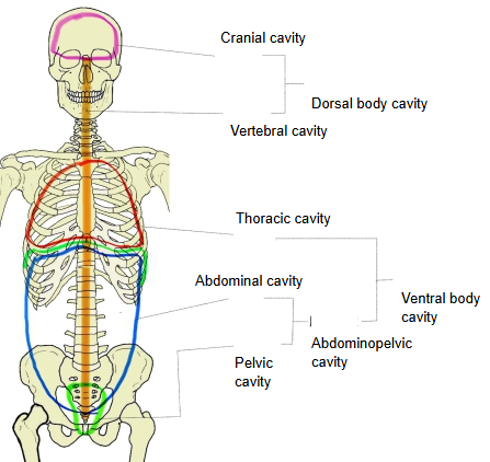
Below is a simple overall diagram of the body cavities. Together with the abdominopelvic cavity below, the thoracic cavity forms the ventral body cavity. (The division into dorsal and ventral cavities is a teaching scheme; it is not part of Terminologia Anatomica.)
Body
cavities
cavities
- Dorsal cavity
- Cranial cavity : main organ: brain; membranes: meninges.
- Vertebral cavity : main organ: spinal cord; membranes: meninges.
- Ventral cavity
- Thoracic cavity
- Right and left pulmonary regions : main organs: the lungs (each in its pleural sac).
- Mediastinum : superior: aortic arch, trachea, oesophagus, thoracic duct and others; inferior: heart (in the pericardial cavity), ascending aorta and others.
- Abdominopelvic cavity
- Abdominal cavity : main organs: digestive organs, spleen, kidneys; membrane: peritoneum.
- Pelvic cavity : main organs: urinary bladder, reproductive organs; membrane: peritoneum.
- Thoracic cavity
 Body cavities (lateral)
 Body cavities (anterior)
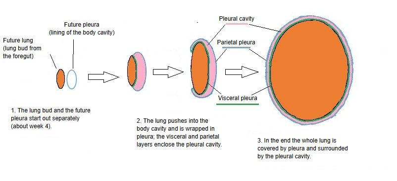
Note: there is also a space called the pleural cavity, but it means a very different space from the thoracic cavity.
The very narrow space between the visceral and parietal pleura, holding a small amount of serous fluid; the thoracic cavity is the whole space inside the chest wall, including the lungs, both pleural sacs and the mediastinum
Reference websites
[Wikipedia]
· Thoracic cavity, Wikipedia.
· Body cavity, Wikipedia.
[Websites with illustrations]
· Body cavities (scheme) (Wikimedia Commons)

· Mediastinum (Kenhub)
Related terms
References and sources
- Further reading: Thoracic cavity, Wikipedia.
- Federative International Programme for Anatomical Terminology (FIPAT). Terminologia Anatomica, 2nd ed. (TA2), 2019.