Thoracic wall (Latin: paries thoracis)
Also called: chest wall
Main components
Bony part: thoracic cage (sternum, the 12 pairs of ribs with their costal cartilages, and the 12 thoracic vertebrae)
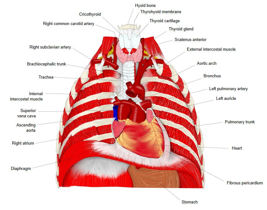
Muscular part: external intercostal muscles, internal intercostal muscles, innermost intercostal muscles, subcostales, transversus thoracis and the diaphragm, which closes the cavity below
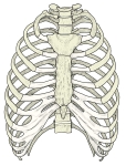 Thoracic cage (anterior view)
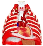 Thoracic wall and its contents
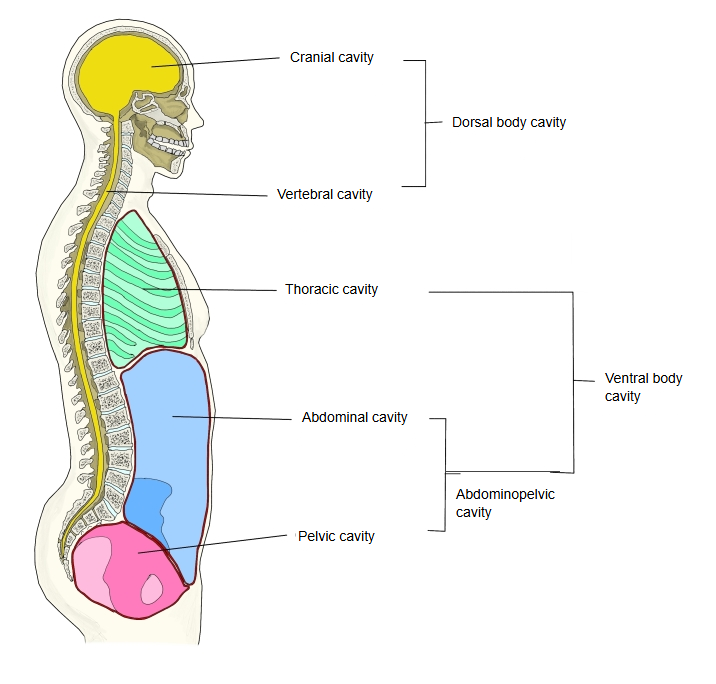
 Body cavities (lateral)
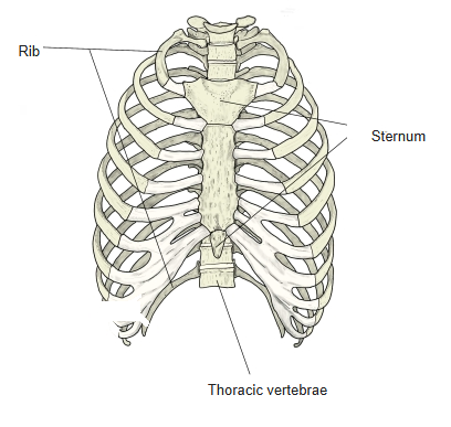
Reference websites
[Wikipedia]
· Thoracic wall, Wikipedia.
· Rib cage, Wikipedia.
[Websites with illustrations]
· Muscles of the thoracic cage (TeachMeAnatomy)
Structure
The bony framework of the thoracic wall is the rib cage; the rest is made up of muscles, skin and fasciae. From outside in, the layers are: the skin, the superficial fascia, the deep fascia, the muscles of the upper limb and back that lie on the cage (such as pectoralis major, serratus anterior or latissimus dorsi, depending on the region), the ribs with the intercostal muscles, and the endothoracic fascia, which lies against the parietal pleura. The intercostal nerves and vessels run in each intercostal space, mainly along the lower border of the rib above. The thoracic wall protects the heart and lungs and changes the volume of the cavity in breathing.
Related terms
- Thorax
- Intercostal space
- External intercostal membrane
- Parietal pleura
- Costal pleura
- Mediastinum
- Sternum
References and sources
- Further reading: Thoracic wall, Wikipedia.
- Further reading: Rib cage, Wikipedia.
- Federative International Programme for Anatomical Terminology (FIPAT). Terminologia Anatomica, 2nd ed. (TA2), 2019.