Thyroid cartilage (Latin: cartilago thyroidea)
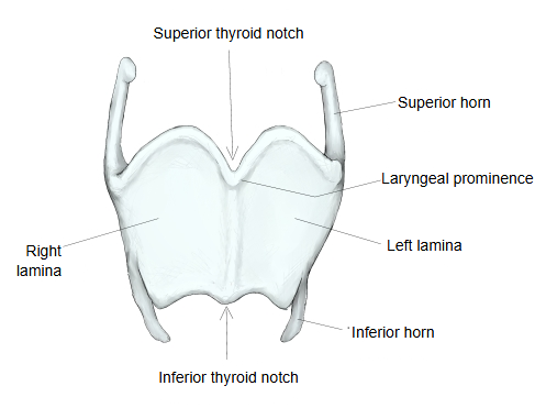
· The right lamina and left lamina are short plates, each with four borders and two surfaces. They join in the midline at about 90° in men and about 120° in women, so the laryngeal prominence (Adam's apple) stands out more in adult men.
Anterior border: short; it is the line where the two laminae meet.
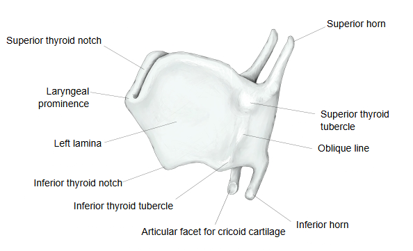
Posterior border: thick and long; it projects upwards and downwards, the upper projection being the superior horn (long, joined to the hyoid bone) and the lower one the inferior horn (short, with a small facet for the cricoid cartilage).
Superior border: has the deep V-shaped superior thyroid notch in the midline. The thyrohyoid membrane joins this border to the hyoid bone.
Inferior border: has the shallow inferior thyroid notch in the midline; the median cricothyroid ligament attaches here.
Inner surface: generally smooth, narrow and slightly concave. Near the midline angle, the stalk of the epiglottis attaches just below the superior thyroid notch, the vestibular ligaments below it, and the vocal ligaments (of the vocal folds) about halfway down the angle, so the cartilage shields the vocal folds.
Outer surface: crossed by the oblique line, which runs from above and behind downwards and forwards, from the superior thyroid tubercle to the inferior thyroid tubercle.
Near the superior thyroid tubercle there is often a small thyroid foramen, through which branches of the superior laryngeal artery and vein and the superior laryngeal nerve pass.
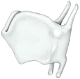 Thyroid cartilage (left side)
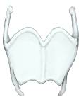 Thyroid cartilage (anterior)
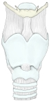 Larynx (anterior)
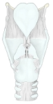 Larynx (posterior)
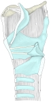 Larynx (median section)
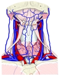 Front of the neck
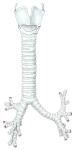 Trachea and bronchi
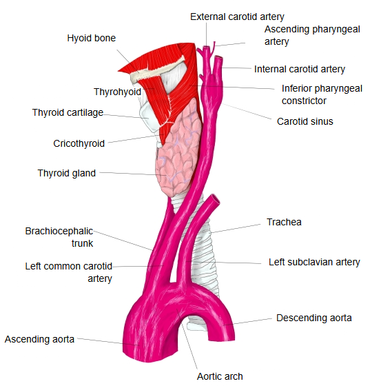
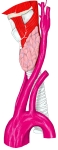 Larynx and great arteries (left side)
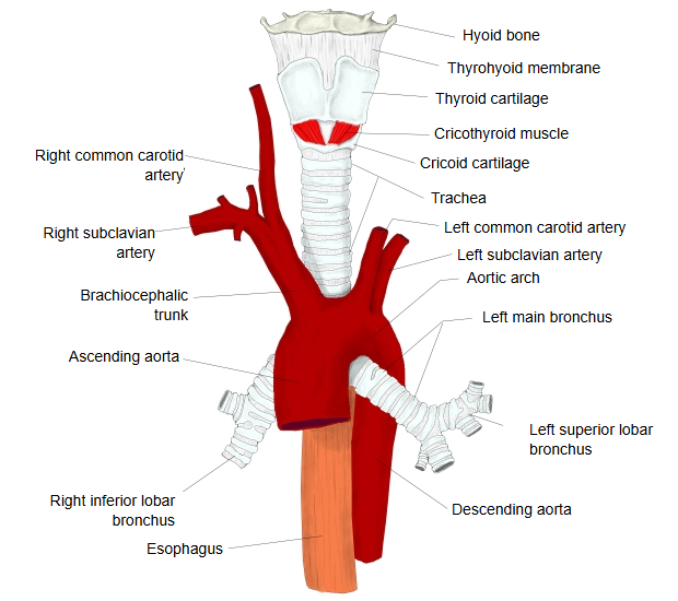
 Larynx, trachea and great vessels
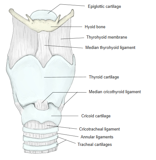
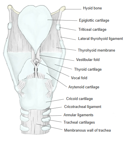
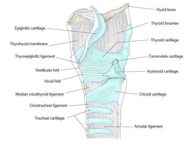
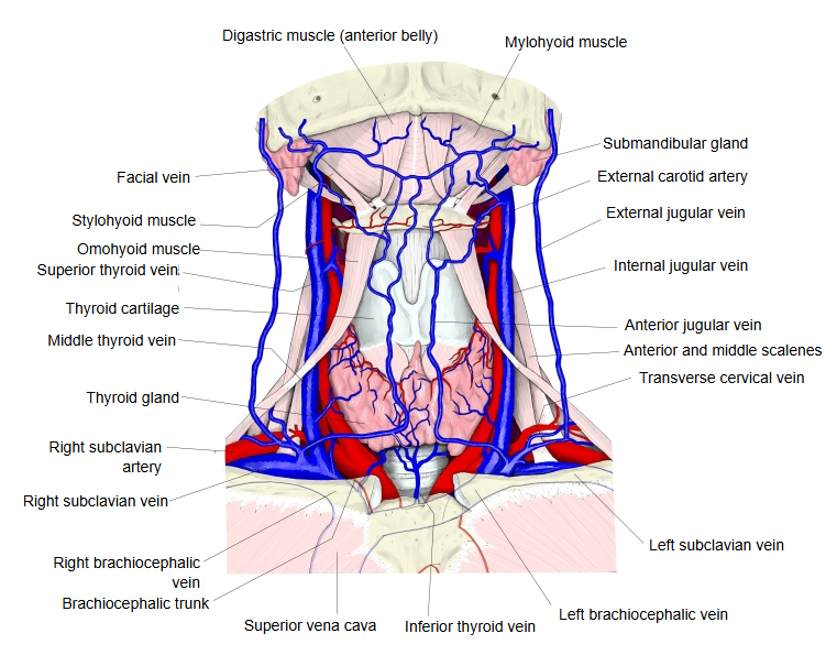
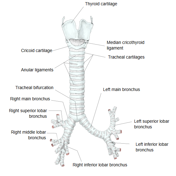
Below is a simple overview of the laryngeal cartilages.
cartilages
- Unpaired
- Thyroid cartilage : the largest; its two laminae form most of the front wall and the laryngeal prominence. (hyaline)
- Cricoid cartilage : the lowest; a complete ring, joined to the first tracheal cartilage by the cricotracheal ligament. (hyaline)
- Epiglottic cartilage : the framework of the epiglottis. (elastic)
- Paired
- Arytenoid cartilage : sits on the upper border of the cricoid lamina; the most important for voice production. (hyaline; vocal process and apex elastic)
- Corniculate cartilage : a very small cartilage on the apex of the arytenoid cartilage. (elastic)
- Cuneiform cartilage : a small, flat cartilage in front of and lateral to the corniculate cartilage; often absent. (elastic)
- Sesamoid cartilages : small inconstant cartilages, sometimes several. The triticeal cartilage in the lateral thyrohyoid ligament is also inconstant.
The thyroid cartilage is hyaline cartilage and unpaired; it is the largest of the laryngeal cartilages, the other unpaired ones being the cricoid and epiglottic cartilages. Its anterior angle, where the right and left laminae meet, can be felt under the skin as the laryngeal prominence, which projects sharply in adult men. The long superior horn runs upwards, backwards and medially and is joined to the hyoid bone by the lateral thyrohyoid ligament; the shorter inferior horn runs downwards and forwards and bears on its inner side the articular facet for the cricoid cartilage, which meets the thyroid articular surface of the cricoid. These are the cricothyroid joints: tilting the thyroid cartilage forwards at them, by the cricothyroid, stretches the vocal folds and raises the pitch of the voice. On the inner surface of the angle, about halfway between the superior thyroid notch and the lower border, the vocal ligaments of the vocal folds attach on either side of the midline. Like other hyaline cartilages of the larynx, the thyroid cartilage often partly ossifies with age.
Reference websites
[Wikipedia]
· Thyroid cartilage, Wikipedia.
· Larynx, Wikipedia.
[Websites with illustrations]
· The larynx (TeachMeAnatomy)
· Thyroid cartilage: structure and function (Kenhub)
· The ligaments of the larynx, antero-lateral view (Gray's Anatomy) (Wikimedia Commons)

Related terms
- Larynx
- Laryngeal prominence
- Right lamina
- Inferior horn of thyroid cartilage
- Inferior thyroid tubercle
- Thyrohyoid membrane
- Cricothyroid
References and sources
- Further reading: Thyroid cartilage, Wikipedia.
- Further reading: Larynx, Wikipedia.
- Federative International Programme for Anatomical Terminology (FIPAT). Terminologia Anatomica, 2nd ed. (TA2), 2019.