Posterior malleolar fold (Latin: plica mallearis posterior)
Also called the posterior mallear fold
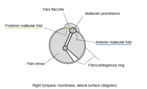
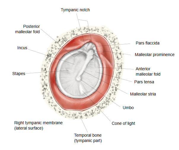
- Anterior malleolar fold : runs obliquely downwards from the greater tympanic spine to the malleolar prominence; the shorter fold.
- Posterior malleolar fold : runs obliquely downwards from the lesser tympanic spine to the malleolar prominence; the longer fold.
Note: the malleolar folds are described above as continuations of the fibrocartilaginous ring (cartilage ring), which is missing at the tympanic notch. Some illustrations instead draw each fold as a separate band, like a ligament, stretched between the tympanic spine and the malleolar prominence; sources differ. Example.
At the border between the pars flaccida and the pars tensa there are arched folds, the malleolar folds, one in front and one behind. The anterior malleolar fold starts at the greater tympanic spine and is short; the posterior malleolar fold starts at the lesser tympanic spine, the posterior end of the tympanic notch, and is long. Both run obliquely downwards to meet at the malleolar prominence over the lateral process of the malleus. A mucosal fold of the same name lies on the inner side of the membrane, in the tympanic cavity.
 Right tympanic membrane (lateral surface)
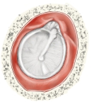 Right tympanic membrane (lateral surface)

Reference websites
[Wikipedia]
· Tympanic membrane, Wikipedia.
[Websites with illustrations]
· Tympanic membrane (diagram) (Epomedicine)
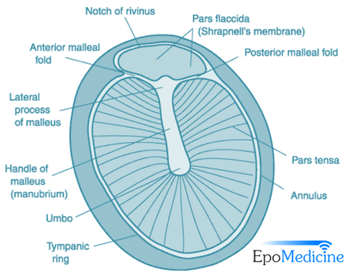
· Tympanic membrane and malleolar folds (image) (Stanford Otology Surgery Atlas)
Related terms
- Tympanic membrane
- Lateral process of malleus
- Malleus
- Middle ear
- Tympanic cavity
- Incudomalleolar joint
References and sources
- Further reading: Tympanic membrane, Wikipedia.
- Federative International Programme for Anatomical Terminology (FIPAT). Terminologia Anatomica, 2nd ed. (TA2), 2019.