Periosteum (Latin: periosteum)
- Overview
- Structure (Outer layer (fibrous layer), Inner layer (osteoblast layer, cambium layer))
- Cellular components
- Periosteum and endosteum compared
- Related terms
Overview
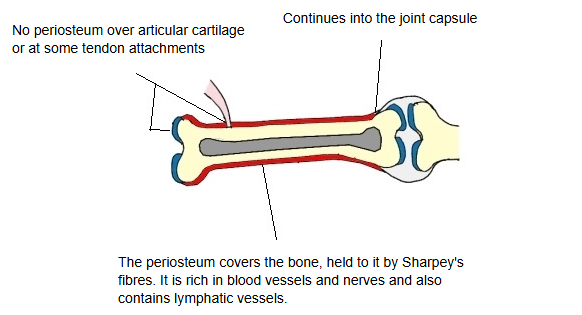
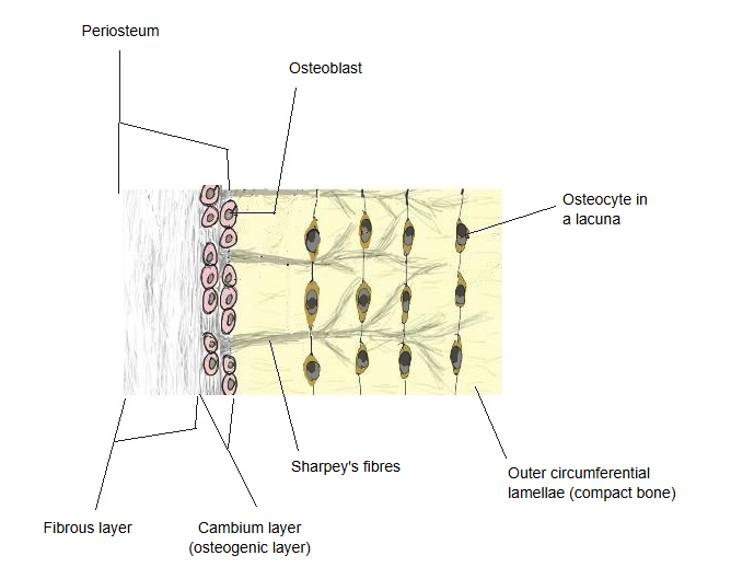
· The periosteum is firmly anchored to the bone by Sharpey's fibres (perforating fibres), bundles of collagen that run from it into the outer lamellae of the bone. At most attachments tendons and ligaments blend with it; only where they are inserted through fibrocartilage is there no periosteum (see above). At a joint it continues into the fibrous layer of the joint capsule.
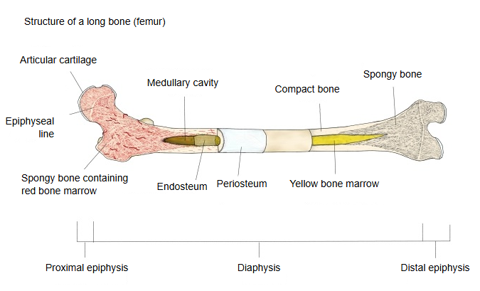
 Periosteum of a long bone
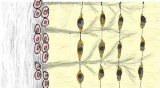 Layers of the periosteum
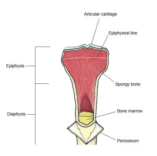
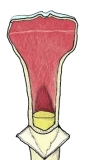 End of a long bone (section)
Structure
of the
periosteum
- Outer layer : the fibrous layer, dense connective tissue made mainly of collagen. Thicker: about 0.05–0.4 mm. Rich in nerves and blood vessels, with some lymphatic vessels.
- Inner layer : the osteoblast layer or cambium layer, rich in cells such as osteoblasts. About 0.01–0.1 mm thick. Its nerves and vessels come from the outer layer; lymphatic vessels are essentially absent.
Outer layer (fibrous layer)
1. Made of dense connective tissue (mainly type I collagen); it links the bone to muscles (through their tendons), ligaments and fascia. Its thickness is about 0.05–0.4 mm, varying with age and site.
2. Rich in blood vessels and nerves, which bring nourishment and sensation to the bone. Its cells are fibroblasts, the endothelial cells of its many vessels and a few immune cells. The vessels enter the bone through small canals such as the perforating canals and the nutrient canal.
Lymphatic vessels are also present, though not many.
3. It has many sensory nerves and takes part in sensing pain, so injuries to bone are painful.
Inner layer (osteoblast layer, cambium layer)
1. Contains mesenchymal stem cells (osteoprogenitor cells), pre-osteoblasts and osteoblasts, the precursors of osteocytes. It is a thin cellular layer (about 0.01–0.1 mm) lying directly on the bone.
2. Takes part in the growth of bone in width (appositional growth: new bone laid down on the surface) and in its repair. It is thicker and more active in children than in adults.
3. After a fracture it becomes very active, and new bone forms from it, making much of the callus.
Cellular components
· Compared with a cell proportion of several tens of percent in the inner layer, the outer layer is thought to have about 10% or less.
| Layer | Cell | Notes | |
|---|---|---|---|
| Inner layer 0.01–0.1 mm | 1 | Mesenchymal stem cells | can become the cells of many kinds of connective tissue; in the periosteum they become the cells below |
| 2 | Pre-osteoblasts | the cells just before osteoblasts; they divide actively and also secrete a little collagen | |
| 3 | Osteoblasts | produce bone matrix and form bone; very active in fracture repair | |
| Outer layer 0.05–0.4 mm | 1 | Fibroblasts | produce collagen fibres; maintain the structure of the fibrous layer |
| 2 | Vascular endothelial cells | line the blood vessels that run through the periosteum; important for nourishment and bone repair | |
| 3 | Immune cells | a few, around the vessels and in the connective tissue; take part in repair and inflammatory responses | |
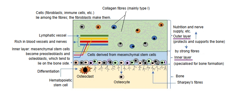
From stem cell to osteocyte
Open question: there may be intermediate cells between mesenchymal stem cells and pre-osteoblasts. English texts usually call the committed precursors osteoprogenitor cells, but the names and the exact stages differ between sources.
Periosteum and endosteum compared
| Item | Periosteum | Endosteum | |
|---|---|---|---|
| 1 | Position | covers the outer surface of the bone | lines the inner surfaces of the bone (the medullary cavity, the trabeculae of spongy bone and the central (Haversian) canals) |
| 2 | Thickness | relatively thick (two layers) | very thin (one layer of bone-lining cells with a little connective tissue) |
| 3 | Structure | two layers: the outer fibrous layer (connective tissue) and the inner osteoblast layer | one layer: osteoblast-like cells with connective tissue |
| 4 | Vessels and nerves | plentiful (nourishment, sensory nerves) | fewer, but the vessels needed for bone remodelling are present |
| 5 | Function | growth in width, repair, blood supply to the outer bone, attachment of tendons and ligaments | bone remodelling (resorption and formation) together with osteoblasts and osteoclasts; forms the boundary with the bone marrow |
| 6 | Where found | the whole outer surface of long, flat and short bones, except the joint surfaces | all inner surfaces of bone: the medullary cavity, the trabeculae of spongy bone, the central canals |
| 7 | Developmental origin | mesenchyme: the perichondrium or the condensed mesenchyme around the developing bone | mesenchymal cells lining the inner surfaces of the developing bone |
Related terms
- Diaphysis
- Epiphysis
- Epiphyseal cartilage
- Endosteum
- Bone marrow
- Bone matrix
- Cambium layer
- Osteoclast
- Perforating canal
- Nutrient canal
- Joint capsule
- Tendon
- Synovial membrane
- Fascia
References and sources
- Further reading: Periosteum, Wikipedia.
- Further reading: Endosteum, Wikipedia.
- Federative International Programme for Anatomical Terminology (FIPAT). Terminologia Anatomica, 2nd ed. (TA2), 2019.