Endosteum (Latin: endosteum)
Overview
Where it is found
1. Inner surface of the medullary cavity: the shaft of a long bone contains the medullary cavity, which holds the bone marrow; the endosteum lines its inner surface.
2. Inner surfaces of the central (Haversian) and perforating (Volkmann's) canals: these are the channels for blood vessels and nerves in compact bone, and the endosteum lines them.
3. Surfaces of the trabeculae of spongy bone: the trabeculae are the thin, intricately branching bony struts of spongy bone, and the endosteum covers their surfaces.
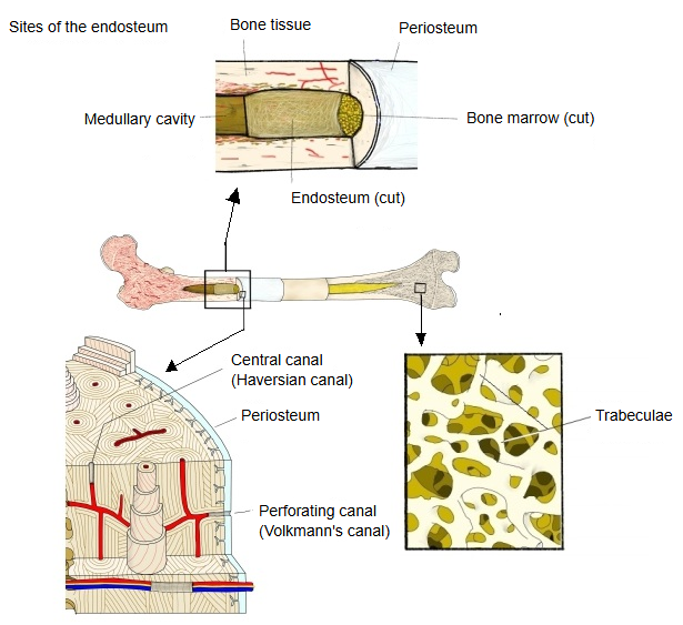
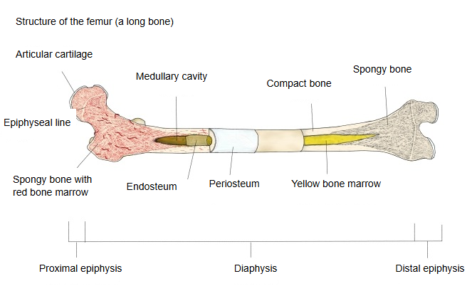
Structure
· It is very thin because, unlike the periosteum, it has no thick fibrous layer: mainly a single layer of bone-lining cells with a little connective tissue. It forms the boundary between bone and bone marrow.
Cells
The following cells are found in the endosteum:
| Cell | Features | Function | |
|---|---|---|---|
| 1 | Osteoblasts | Bone-forming cells; cuboidal, lined up along the bone surface | Lay down new bone matrix (osteoid) and help mineralise it; the centre of bone formation |
| 2 | Osteoprogenitor cells (mesenchymal stem cells) | Undifferentiated or partly differentiated cells | Become osteoblasts; called on during remodelling; the source of bone repair and regeneration |
| 3 | Bone-lining cells | Flattened, resting osteoblasts | Cover quiet bone surfaces; help control bone resorption and formation; maintain and protect the bone |
| 4 | Osteoclasts | Large cells with many nuclei that resorb bone | Break down bone matrix and release calcium and phosphate; the resorbing side of remodelling |
| 5 | Stromal cells | Lie in the thin connective tissue | Support osteoblasts and osteoclasts; take part in the niche for blood-forming stem cells |
Comparison of the periosteum and the endosteum
| Feature | Periosteum | Endosteum | |
|---|---|---|---|
| 1 | Position | Covers the outer surface of bone (except the articular cartilage) | Lines the inner surfaces of bone (medullary cavity, trabeculae of spongy bone, central canals) |
| 2 | Thickness | Relatively thick (two layers) | Very thin (one layer of cells with a little connective tissue) |
| 3 | Structure | Two layers: outer fibrous layer and inner cellular (osteogenic) layer | One layer: osteoblast-like (bone-lining) cells with connective tissue |
| 4 | Vessels and nerves | Rich in vessels and sensory nerves | Fewer, but has the vessels needed for remodelling |
| 5 | Function | Adds bone to the outer surface (growth in width); fracture repair; blood supply to the outer bone | Remodels bone from the inside (resorption and formation); boundary with the bone marrow |
| 6 | Where found | The whole outer surface of long, flat and short bones | All inner surfaces of bone: medullary cavity, trabeculae, central canals |
| 7 | Origin | Mesenchyme around the developing bone | Mesenchyme around the developing bone (cells lining the inner surfaces) |
Related terms
- Diaphysis
- Epiphysis
- Epiphyseal cartilage
- Periosteum
- Bone marrow
- Bone matrix
- Cambium layer
- Perforating canal
- Osteoclast
- Nutrient canal
- Articular cartilage
- Proximal epiphysis of femur
- Femur
References and sources
- Further reading: Endosteum, Wikipedia.
- Further reading: Periosteum, Wikipedia.
- Federative International Programme for Anatomical Terminology (FIPAT). Terminologia Anatomica, 2nd ed. (TA2), 2019.