Cisterna chyli (Latin: cisterna chyli)

Also called the receptaculum chyli or the chyle cistern

Overview

The cisterna chyli is part of the lymphatic system: a sac-like structure that forms the beginning of the thoracic duct.
Lymphatic system: lymph capillaries, lymph vessels, lymph nodes, lymphatic trunks and ductsThoracic duct: a lymph vessel about 38–45 cm long that rises along the vertebral column

· Size: a few centimetres long and about 1–2 cm wide (sources differ a good deal).

· Shape: an elongated, flattened sac, or a triangular (pyramid-shaped) one.

· Position: behind the peritoneum, on the front of the bodies of the first and second lumbar vertebrae, just to the right of and behind the abdominal aorta, near the origin of the superior mesenteric artery.

· It does not always form a clear swelling: in many people the trunks join without a distinct sac, or form a small plexus instead.

Relations: in front, the right crus of the diaphragm and the abdominal aorta partly overlap it; behind lie the bodies of the first and second lumbar vertebrae; above, it narrows into the thoracic duct, which passes through the aortic hiatus at about T12.

Sources differ on its size and exact level (from T12 to L2), and on how often a distinct sac is found.
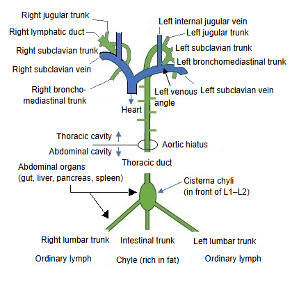
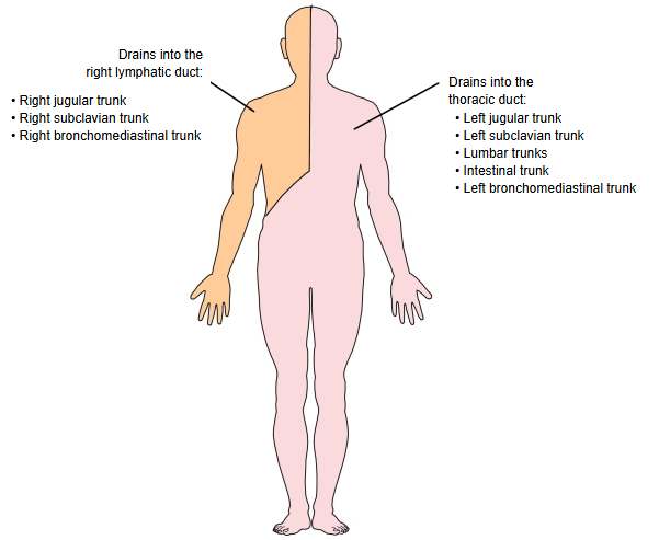
The lymph of the right and left lumbar trunks and of the intestinal trunk flows into the cisterna chyli.
Lumbar trunks: collect the lymph of the lower limbs, the pelvis and the lower abdominal wallIntestinal trunk: collects the lymph (and fat-rich chyle) of the small intestine and the other organs of the abdominal cavity
Diagram of the lymph trunks joining the thoracic duct and the right lymphatic duct, with the cisterna chyli, aortic hiatus and venous angles labelled.
Lymph trunks and ducts (diagram) (click to enlarge)
A scheme of the lymph trunks of each side joining the right lymphatic duct or, via the cisterna chyli, the thoracic duct, with the size of each duct and the share of the body's lymph it carries.
Lymph trunks and ducts (scheme) (click to enlarge)

The path of chyle

villus
Central lactealin the centre of the villus; joins the lymph vessels below
submucosa
Intestinal lymph vesselsshort course; soon leave the gut wall
mesentery
Mesenteric lymph vesselspass through the mesenteric lymph nodes
abdomen
Intestinal trunkabout 1.5–4 cm; soon enters the cisterna chyli
Cisterna chyliin front of L1–L2; chyle mixes with the lymph of the lumbar trunks
through the aortic hiatus into the thorax
Thoracic duct38–45 cm; rises behind and to the right of the aorta; arches forward at about C6–C7
Superior vena cava
Heart

Chyle is the milky, fat-rich lymph of the intestine; the thoracic duct carries it together with ordinary lymph.
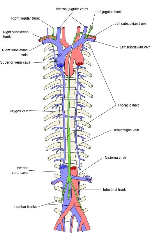
  • The thoracic duct in green rising from the cisterna chyli between the azygos and hemiazygos veins, with the venae cavae and lymph trunks labelled.Thoracic duct and the azygos system
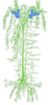
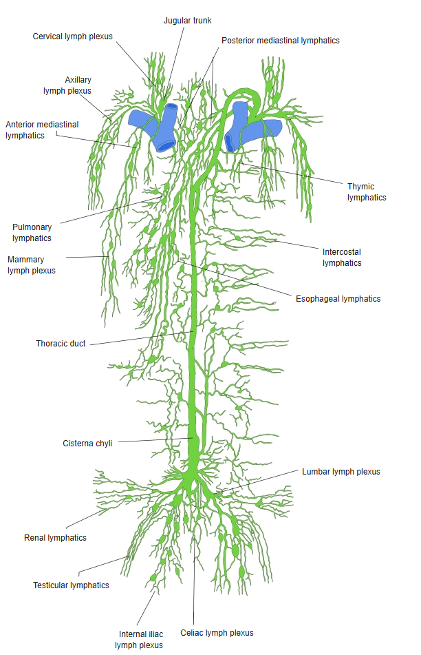
  • The lymph vessels and lymph trunks of the trunk draining into the cisterna chyli and the thoracic duct.Lymph vessels of the trunk
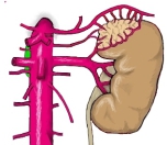
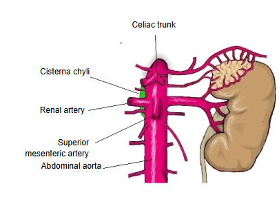
  • The abdominal aorta with the cisterna chyli in green beside it, and the celiac trunk, renal artery and superior mesenteric artery labelled.Cisterna chyli and the abdominal aorta
Reference websites

[Wikipedia]

· Cisterna chyli, Wikipedia.

· Thoracic duct, Wikipedia.

[Websites with illustrations]

· Abdominal lymph nodes, from the iliac nodes to the cisterna chyli (Shutterstock)

· Thoracic duct, cisterna chyli, lumbar and intestinal trunks and the abdominal and pelvic lymph nodes (Clinical Imaging)

References and sources

  1. Further reading: Cisterna chyli, Wikipedia.
  2. Further reading: Thoracic duct, Wikipedia.
  3. Federative International Programme for Anatomical Terminology (FIPAT). Terminologia Anatomica, 2nd ed. (TA2), 2019.