Utriculosaccular duct (Latin: ductus utriculosaccularis)
Also called the utriculosaccular canal
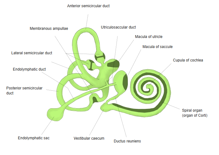
The utriculosaccular duct is the fine duct, in the middle part of the membranous labyrinth, that joins the saccule and the utricle.
= the part lying in the vestibule of the bony labyrinthBoth carry sensory epithelial cells (hair cells)
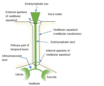
From the utriculosaccular duct the endolymphatic duct arises; it runs through the vestibular aqueduct towards the cranial cavity and ends as the endolymphatic sac.
= the bony canal of the vestibule (in the petrous part of the temporal bone)Contains endolymph; a very narrow ductLies between the two layers of the dura mater
Two sacs of the vestibule
- Saccule : round and flattened, about 3 mm long and 2 mm wide; lies in the spherical recess. Its macula detects linear acceleration in the vertical plane.
- Utriculosaccular duct : joins the two sacs and gives off the endolymphatic duct (which passes through the vestibular aqueduct and ends in the endolymphatic sac).
- Utricle : an elongated oval sac about 5–6 mm long; lies in the elliptical recess. Its macula detects linear acceleration in the horizontal plane. The semicircular ducts open into it.
· Sources differ in how they describe the junction: some treat it as one duct between the utricle and the saccule that gives off the endolymphatic duct, others as a short utricular duct and a saccular duct that unite to form the endolymphatic duct.
 Membranous labyrinth
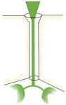 Utriculosaccular duct to endolymphatic sac (schematic)
Reference websites
[Wikipedia]
· Membranous labyrinth, Wikipedia.
· Endolymphatic duct, Wikipedia.
[Websites with illustrations]
· Inner ear with the utriculosaccular and endolymphatic ducts (veterinary textbook figure) (Veterian Key)

Related terms
- Elliptical recess
- Macula of utricle
- Endolymphatic duct
- Endolymphatic sac
- Macula of saccule
- Vestibular maculae
- Utricle
- Saccule
- Membranous labyrinth
References and sources
- Further reading: Membranous labyrinth, Wikipedia.
- Further reading: Endolymphatic duct, Wikipedia.
- Federative International Programme for Anatomical Terminology (FIPAT). Terminologia Anatomica, 2nd ed. (TA2), 2019.