Juxtaglomerular cells
Also called granular cells or JG cells
- Overview
- Structure of the arterial wall
- Reference websites
- (Wikipedia, websites with illustrations)
- Related terms
Overview
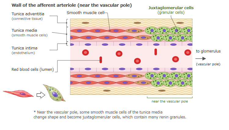
· In other words, juxtaglomerular cells are ordinary smooth muscle cells that have changed into a special form containing granules (which contain renin).
· They are also called granular cells. Note that cells called granule or granular cells also exist elsewhere, for example in the cerebellum and the skin; those are different cells.
· They lie mainly in the wall of the afferent arteriole, and a few in the wall of the efferent arteriole. The name comes from their position next to (juxta-) the glomerulus.
· Their cytoplasm holds renin-containing secretory granules, which give the cells a granular look in stained sections. Their nuclei are rounded rather than elongated.
· Renin is released when the pressure of the blood flowing into the kidney falls (sensed by stretch receptors in the vessel wall), when the macula densa detects a fall in the NaCl concentration of the tubular fluid, and under stimulation by sympathetic nerves (beta-1 receptors).
· Renin acts as an enzyme that converts angiotensinogen into angiotensin I. Through the renin-angiotensin-aldosterone system it raises blood pressure and body fluid volume.
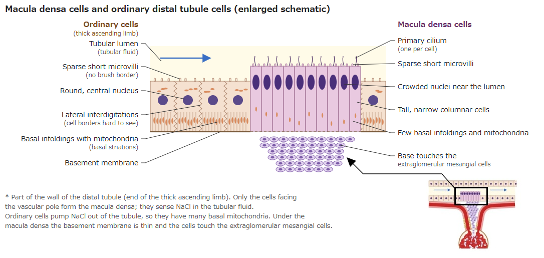
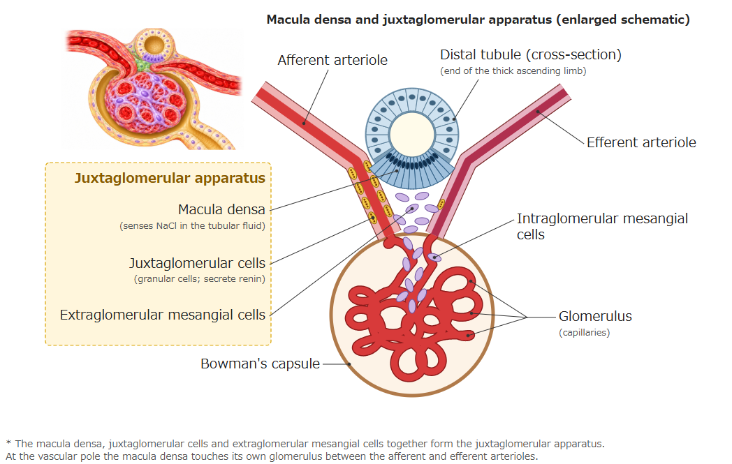
Structure of the arterial wall
The wall of an ordinary afferent arteriole has the three layers shown below.
- Tunica intima
- Endothelial cells : a single layer of flat cells in contact with the blood.
- Tunica media
- Smooth muscle cells : one or two layers; they adjust the width of the vessel.
- Juxtaglomerular cells : near the vascular pole, smooth muscle cells of the media change shape; they contain renin granules.
- Smooth muscle cells : one or two layers; they adjust the width of the vessel.
- Tunica adventitia
- Connective tissue : a thin layer of fibres and fibroblasts.
Reference websites
[Wikipedia]
· Juxtaglomerular cell, Wikipedia.
· Juxtaglomerular apparatus, Wikipedia.
· Renin, Wikipedia.
[Websites with illustrations]
· Juxtaglomerular cells (micrograph) (SIU School of Medicine histology)
· Tubules and collecting system (8) (Digital Histology)
· Tubules and collecting system (7) (Digital Histology)
Related terms
- Juxtaglomerular apparatus
- Macula densa
- Vascular pole
- Distal tubule
- Kidney
- Renal artery
- Interlobular arteries of kidney (cortical radiate arteries)
- Renal parenchyma
- Urinary system
References and sources
- Further reading: Juxtaglomerular cell, Wikipedia.
- Further reading: Juxtaglomerular apparatus, Wikipedia.
- Further reading: Renin, Wikipedia.
- Federative International Programme for Anatomical Terminology (FIPAT). Terminologia Anatomica, 2nd ed. (TA2), 2019.