Tunica adventitia (Latin: tunica adventitia)
Also called the adventitia; in blood vessels, the tunica externa
- Overview
- Where it is found
- Serosa and adventitia compared
- Reference websites
- (Wikipedia, websites with illustrations)
- Where it is found
- Related terms
Overview
Note: the epicardium and the perimetrium are called the outer layers of the heart and the uterus, but both are serous membranes, not adventitia.
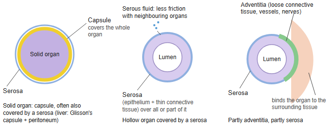
Where part of the circumference of an organ (as in the ascending and descending colon) or all of it (as in the oesophagus) has no serosa, the organ is wrapped only in connective tissue with no clear boundary; this covering is the adventitia. Organs at the back of the abdomen are wrapped in adventitia of loose connective tissue.
The table below lists the cells found in the connective tissue of the adventitia.
(Cells of the vessels and nerves are not included.)
| Cell | Group | Main role | |
|---|---|---|---|
| 1 | Fibroblasts | Connective tissue cells | Make collagen fibres and elastic fibres |
| 2 | Fibrocytes | Connective tissue cells | Maintain the connective tissue (resting form of the fibroblast) |
| 3 | Macrophages | Immune cells | Remove foreign material; immune defence |
| 4 | Mast cells | Immune cells | Inflammation; control of vessel permeability |
| 5 | Lymphocytes | Immune cells | Immune surveillance (few) |
| 6 | Plasma cells | Immune cells | Make antibodies (more numerous in inflammation) |
around organs
and tubes
- Serosa : structure: a simple squamous epithelium (mesothelium) on a thin layer of connective tissue; secretes serous fluid that reduces friction. It covers organs that face a body cavity: the pleura, pericardium and peritoneum (each organ's part has its own name), e.g. the small and large intestines (in part).
- Adventitia : structure: loose connective tissue with vessels and nerves; anchors the organ to the surrounding tissue, with no clear boundary. E.g. oesophagus, duodenum, parts of the large intestine, large blood vessels, trachea and bronchi.
- Capsule : the outer layer that is part of a solid organ, so not the same as an "organ + membrane" arrangement; it can be covered further by a serosa or an adventitia.
- Not covered by these
- Open to the outside : oral cavity, nasal cavity, pharynx, larynx, external acoustic meatus, anal canal.
- Central nervous system : brain and spinal cord: covered by the meninges.
- Others : bone: periosteum; muscle: fascia; skin: epidermis and dermis.
Capsule: a membrane (dense connective tissue) that is part of a solid organ; it may be covered further by a serosa or an adventitia.
Serosa: "cell layer + connective tissue layer"; covers organs facing a body cavity and secretes serous fluid that reduces friction.
Adventitia: "loose connective tissue + vessels and nerves"; anchors the organ to the surrounding tissue.
Where it is found
| Digestive tract: adventitia as the outermost layer | ||
| 1 | Oesophagus (cervical and thoracic parts) | Lies outside the body cavities, so it has no mesothelium (serosa). Abdominal part: a short segment inside the abdominal cavity; basically adventitia, but serosa (peritoneum) covers part of its front. |
| 2 | Duodenum | The first part is almost entirely covered by serosa; the rest is retroperitoneal, with serosa on its front and adventitia on its back. |
| 3 | Ascending colon | Mostly adventitia (on the back), but the front is covered by serosa. |
| 4 | Descending colon | |
| 5 | Rectum | Above the peritoneal reflection the front is covered by serosa; below it the rectum has only an adventitia. |
| Respiratory system (tubes): adventitia as the outermost layer | ||
| 1 | Trachea | Covered all round. |
| 2 | Main and segmental bronchi | Inside the lung, the adventitia becomes continuous with the surrounding connective tissue. |
| Circulatory system (blood vessels): adventitia (tunica externa) | ||
| 1 | Arteries and veins | The vessel wall has three coats, intima, media and adventitia → the outermost is always the adventitia (e.g. the aorta). |
| Urinary system (tubes): adventitia as the outermost layer | ||
| 1 | Ureter | Covered by adventitia along its whole length. |
| 2 | Urethra | |
| Genital system (tubes): adventitia as the outermost layer | ||
| 1 | Ductus deferens | Covered all round by adventitia. Where the peritoneum lies next to it, part of the adventitia may seem to touch the peritoneum. |
| 2 | Uterine tube (only a tiny part) | Basically covered all round by serosa. Only very small regions, such as the intramural part that runs through the wall of the uterus, lie away from the peritoneum and have an adventitia-like covering. |
Serosa and adventitia compared
| Feature | Serosa | Adventitia | |
|---|---|---|---|
| 1 | Definition | Membrane covering the outermost layer of organs inside a body cavity (e.g. the visceral peritoneum) | Connective tissue forming the outermost layer of organs outside a body cavity |
| 2 | Position of the organs | Inside a body cavity (abdominal, thoracic, pericardial) | Outside a body cavity, or fixed to the body wall |
| 3 | Epithelium | Present (mesothelium) | Absent |
| 4 | Surface epithelium | Simple squamous epithelium (mesothelium) | None |
| 5 | Main components | Mesothelium + loose connective tissue | Loose to dense connective tissue |
| 6 | Histological features | Smooth and slippery | Continuous with and fixed to the surrounding tissue |
| 7 | Function | Reduces friction; lets the organ slide; secretes serous fluid | Fixes and supports the organ |
| 8 | Relation to cavity fluid | Lubricated by serous fluid | None |
| 9 | Vessels and nerves | In the connective tissue layer | Often plentiful |
| 10 | Typical examples | Stomach, small intestine, transverse colon | Oesophagus, rectum, great vessels; the back of retroperitoneal organs such as the pancreas |
Reference websites
[Wikipedia]
· Adventitia, Wikipedia.
· Serous membrane, Wikipedia.
[Websites with illustrations]
· Overview of the digestive system (layers of the alimentary canal) (OpenStax Anatomy and Physiology 2e)
· Structure and function of blood vessels (OpenStax Anatomy and Physiology 2e)
Where it is found
- Digestive tract: the cervical and thoracic esophagus; the back of the duodenum, ascending and descending colon; the rectum below the peritoneal reflection.
- Airways: the trachea and the main and segmental bronchi outside the lung.
- Blood vessels: arteries and veins have three coats (intima, media, adventitia); the adventitia is always the outer one.
- Urinary and genital ducts: the ureter, the urethra and the ductus deferens.
Related terms
References and sources
- Further reading: Adventitia, Wikipedia.
- Further reading: Serous membrane, Wikipedia.
- Federative International Programme for Anatomical Terminology (FIPAT). Terminologia Anatomica, 2nd ed. (TA2), 2019.