Retrorectal space (Latin: spatium retrorectale)
Also called the presacral space (nearly the same)
- Overview
- Boundaries
- Contents
- Reference websites
- (Wikipedia, websites with illustrations)
- Related terms
Overview
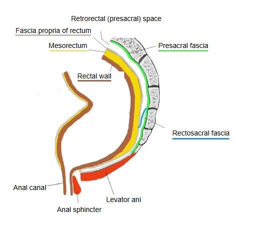
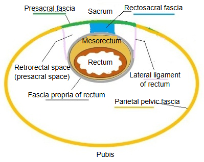
· It lies behind the rectum and its mesorectum and in front of the sacrum and coccyx. Normally it is only a thin layer of loose areolar tissue with a little fat; it opens up as a space only when it is dissected or filled by disease.
· Surgeons separate the rectum from the sacrum through it in rectal cancer surgery, keeping the fascia around the mesorectum intact. Lower down, the rectosacral fascia crosses it and divides it into an upper and a lower part.
Boundaries
What bounds the retrorectal space, in a short table:
| Side | Notes | |
|---|---|---|
| 1 | In front | The rectum with its mesorectum (fatty tissue), wrapped in the fascia propria of the rectum |
| 2 | Behind | The presacral fascia covering the front of the sacrum and coccyx |
| 3 | Above | The level where the peritoneum turns back (the peritoneal reflection); here it continues into the retroperitoneal tissue in front of the promontory, with the fascia around the ureters and hypogastric nerves |
| 4 | Below | The pelvic floor, where the levator ani and the fasciae of the rectum and sacrum meet |
| 5 | At the sides | The lateral ligaments of the rectum and the pelvic side walls |
Contents
What is found in the retrorectal space, in a short list:
| Connective tissue | 1 | Fibrous tissue | Mostly loose connective tissue. |
| 2 | Adipose tissue | Between the fibres of the loose connective tissue; sources differ on the amount, from very little to moderate. | |
| Vessels | 1 | Median sacral artery and vein | Run up and down the midline of the sacrum. |
| 2 | Lateral sacral arteries and veins | Run up and down near the sacral foramina on each side. | |
| 3 | Sacral (presacral) venous plexus | A network of veins spread over the front of the sacrum. | |
| Nerves | 1 | Hypogastric nerves | Sympathetic nerves that descend from the pelvic inlet at the back and sides of the space. |
| 2 | Pelvic splanchnic nerves | Parasympathetic nerves from the 2nd to 4th sacral nerves, coming forward through the anterior sacral foramina. | |
| 3 | Inferior hypogastric (pelvic) plexus | The large network formed where the nerves above meet. | |
| 4 | Sympathetic trunk | A pair of nerve trunks descending on the front of the sacrum. | |
| Others | 1 | Sacral lymph nodes | Lymph nodes along the vessels. |
Sources differ on the extent of this space. In the broad sense (all the tissue between the rectum and the sacrum, as used for retrorectal tumours) it includes the vessels and nerves listed above. In the narrow surgical sense it is only the nearly bloodless plane between the fascia propria of the rectum and the presacral fascia; the sacral vessels, the venous plexus and the sympathetic trunk then lie behind the presacral fascia, and the pelvic nerves at its sides.
Reference websites
[Wikipedia]
· Total mesorectal excision, Wikipedia.
· Pelvic fascia, Wikipedia.
[Websites with illustrations]
· Spaces of the female pelvis, including the presacral (retrorectal) space (cross-section) (Abdominal Key)
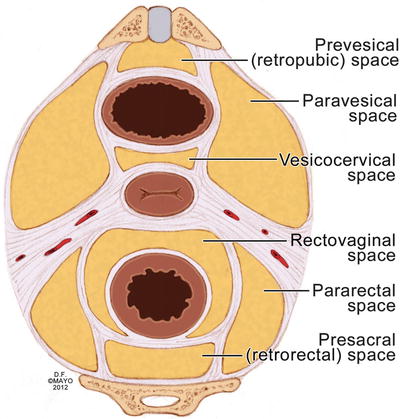
· Pelvic spaces and ligaments, with the presacral (retrorectal) space (cross-section) (Springer)

Related terms
- Fascia propria of rectum
- Rectosacral fascia
- Transverse folds of rectum
- Rectococcygeus
- Presacral space
- Presacral fascia
- Mesorectum
- Rectum
- Sacrum
- Parietal pelvic fascia
- Sacral venous plexus
References and sources
- Further reading: Total mesorectal excision, Wikipedia.
- Further reading: Pelvic fascia, Wikipedia.
- Federative International Programme for Anatomical Terminology (FIPAT). Terminologia Anatomica, 2nd ed. (TA2), 2019.