Crypt (Latin: crypta)
Overview
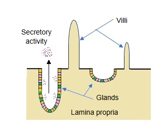
A crypt is a tubular or pit-like structure formed where the epithelium sinks inwards (towards its base) into the connective tissue beneath it.
Epithelium: the layer of cells covering the outer surface of the body and the surfaces of the body cavities and hollow organs
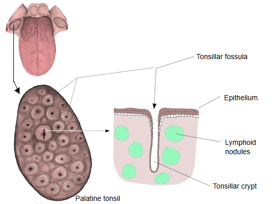
· The deepest crypts: the crypts of the palatine tonsil, several millimetres deep (sometimes 3–10 mm) and branched.
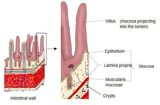
· Among the shallowest: the crypts of Lieberkühn (intestinal crypts) of the small intestine, about 100–200 µm, only a fraction of a millimetre.
Crypts can be divided into two broad groups: those that secrete (glands) and those that do not.
Most crypts; found in the stomach, small intestine and large intestineThe tonsils and others
[ List of crypts ] (this list may not be complete)
| Secretory | Name | Site | As a gland | |
| 1 | Crypts of Lieberkühn (intestinal crypts) | Mucosa of the small intestine | Secretory gland. Function: secretion of digestive juice and mucus; renewal of the epithelium by its stem cells. Contains Paneth cells, goblet cells and enteroendocrine cells; opens between the villi. | |
| 2 | Crypts of Lieberkühn | Mucosa of the large intestine | Secretory gland: long, straight and rich in goblet cells; secretes mucus and helps absorb water and electrolytes; more specialized for secretion than in the small intestine; no villi. | |
| 3 | Gastric glands (gastric pit + glandular part) | Mucosa of the stomach | Secretory gland. Function: secretion of gastric acid, pepsinogen and mucus. Strictly, the gastric pits are the crypt-like part. | |
| Non-secretory | 1 | Crypts of the palatine tonsil (tonsillar crypts) | Oropharynx | Palatine tonsil. Function: trapping antigens, immune responses. Deep and branched; shed cells and debris easily collect in them. |
| 2 | Crypts of the lingual tonsil | Root of the tongue | Lingual tonsil. Function: local immunity, antigen presentation. Short and usually unbranched; mucous glands are separate and open near them. | |
| 3 | Crypts of the pharyngeal tonsil | Nasopharynx | Pharyngeal tonsil: folds and a few shallow crypts. Function: trapping inhaled antigens; well developed in children. | |
| 4 | Dips of the epithelium over the aggregated lymphoid nodules | End of the ileum and elsewhere | Peyer's patches. Function: gut immunity, uptake of antigens. M cells take up antigens, but these are not glands. | |
| 5 | Dips of the epithelium around lymphoid follicles | Upper respiratory tract, gastrointestinal tract | Mucosa-associated lymphoid tissue. Function: immune surveillance; sometimes described as crypt-like structures. |
 Intestinal wall
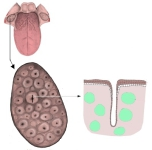 Palatine tonsil
Reference websites
[Wikipedia]
· Intestinal gland, Wikipedia.
· Tonsil, Wikipedia.
[Websites with illustrations]
· The crypt-villus unit of the small intestine and its cells (figure) (Nature Cell Biology)

· Crypts of the small intestine and colon (figure) (International Journal of Molecular Sciences (MDPI))

· Villus and crypt base with stem cells and Paneth cells (figure) (Cell Regeneration)

Related terms
- Intestinal crypts
- Tonsillar crypts
- Villi
- Gastric pits
- Palatine tonsil
- Paneth cell
- Aggregated lymphoid nodules
References and sources
- Further reading: Intestinal gland, Wikipedia.
- Further reading: Tonsil, Wikipedia.
- Federative International Programme for Anatomical Terminology (FIPAT). Terminologia Anatomica, 2nd ed. (TA2), 2019.