Palatine tonsil (Latin: tonsilla palatina)
Also called the faucial tonsil or the "the tonsil" or the plural palatine tonsils
- Overview
- Vessels and nerves
- Reference websites
- (Wikipedia, websites with illustrations)
- Where it is found
- Related terms
Overview
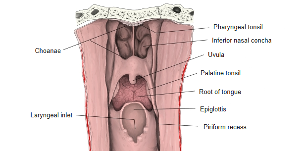
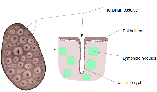
- Palatine tonsil : paired; the largest tonsil, oval, in the tonsillar sinus of the fauces; its surface shows the tonsillar pits.
- Lingual tonsil : on the surface of the back third of the tongue (root of the tongue); contains dense lymphoid nodules.
- Tubal tonsil : paired; around the pharyngeal opening of the auditory tube, in the torus tubarius.
- Pharyngeal tonsil : in the upper part of the back wall of the nasopharynx (the vault of the pharynx).
The shape of the palatine tonsil differs from person to person: it may bulge out of the tonsillar sinus into the cavity of the pharynx, it may be quite flat, or its centre may be sunk with a raised rim around it.
Like other mucosa-associated lymphoid tissue, the palatine tonsil takes part in the local immune response to microorganisms that enter through the mouth and nose. Inflammation of the palatine tonsils is tonsillitis; pus outside the capsule is a peritonsillar abscess (quinsy).
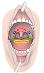 Tonsils (from the front)
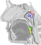 Tonsils (median section)
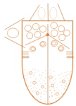 Tongue (schematic)
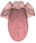 Tongue and surroundings
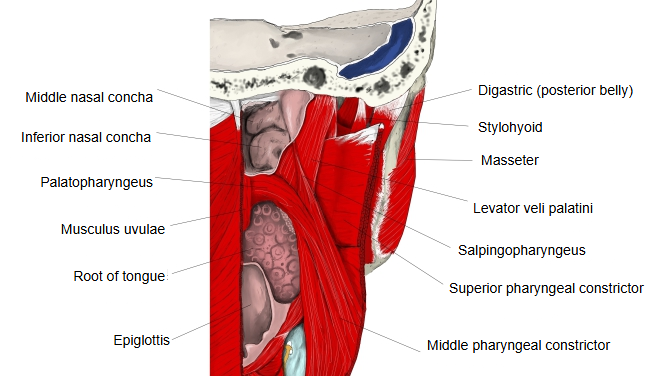
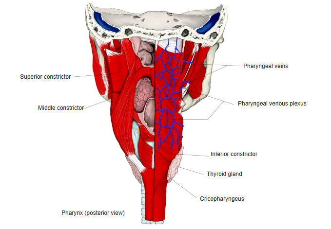
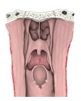 Pharynx (opened from behind)
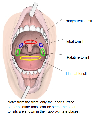
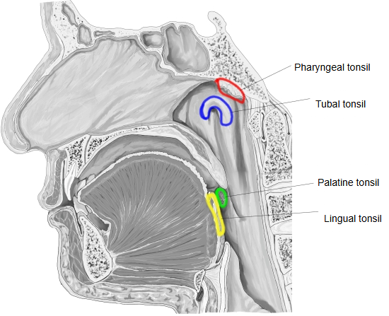
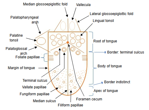
· The medial surface faces the oropharynx and is covered by non-keratinized stratified squamous epithelium; it shows the openings of about 10–20 tonsillar pits, which lead into the deep tonsillar crypts. Lymphoid nodules with germinal centres lie beneath the epithelium and along the crypts (sources differ on the number of crypts). The supratonsillar fossa is a remnant of the second pharyngeal pouch.
Relations
· The lateral surface of the palatine tonsil is covered by a fibrous capsule, which sends septa into the tonsil and faces the palatopharyngeus and the superior pharyngeal constrictor, which (with the styloglossus) form the tonsillar bed.
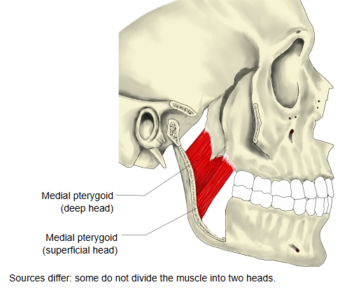
· Outside the wall of the pharynx lies the medial pterygoid; the space behind it is filled with loose tissue that carries vessels and nerves.
(The important vessel closest to the tonsil is the facial artery, especially where it loops.)
 Palatopharyngeus
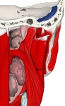 Muscles of the pharynx (from behind)
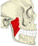 Medial pterygoid
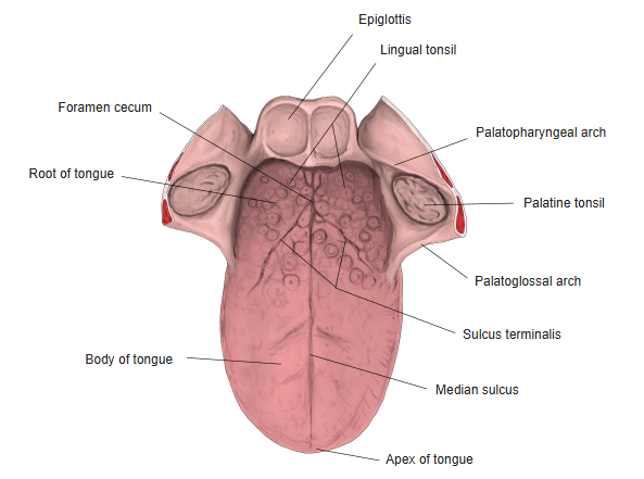 Tongue and its surroundings
- Pharynx (opened from behind)
Vessels and nerves
The vessels and nerves of the palatine tonsil are:
Arteries
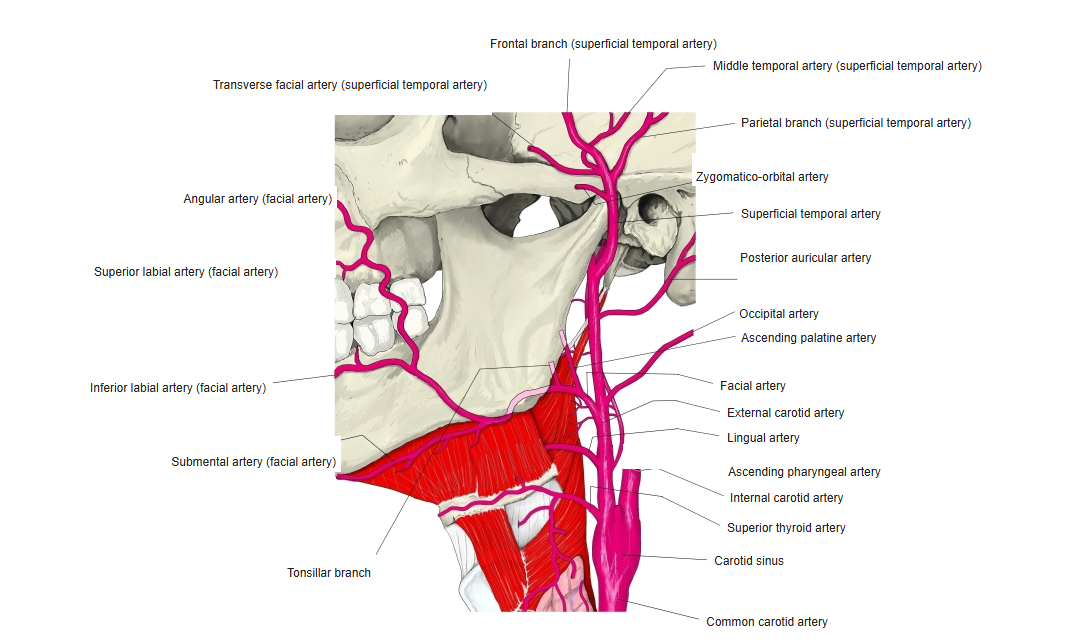
· Ascending palatine artery and tonsillar branch from the facial artery (the tonsillar branch is the main supply).
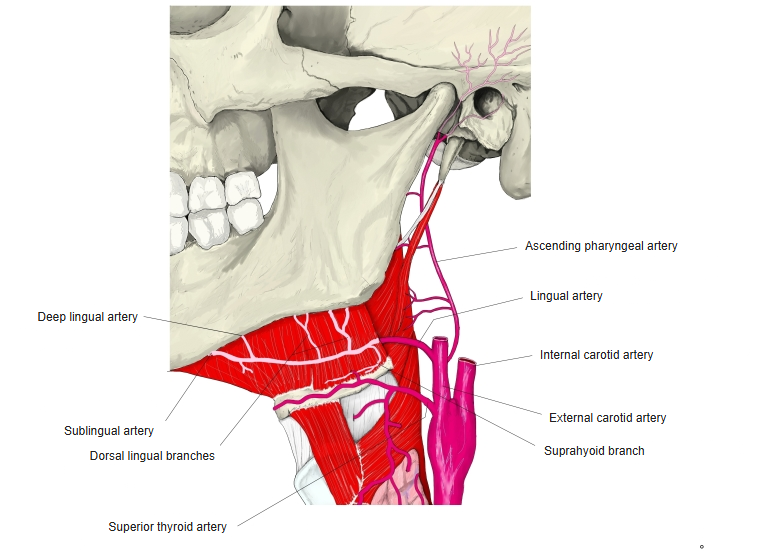
· The ascending pharyngeal artery from the external carotid artery, and the dorsal lingual branches of the lingual artery.
The facial artery is the important vessel closest to the tonsil, especially where it loops; its ascending palatine artery and tonsillar branch are the most closely related. The internal carotid artery and the internal jugular vein lie deeper, behind and lateral to the tonsil (sources give about 1 to 2.5 cm), and the external carotid artery lies farther out. The ascending pharyngeal artery lies behind the tonsil.
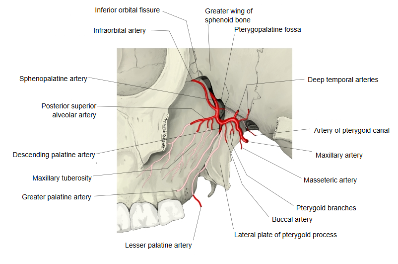
Besides these arteries, English Wikipedia also names the lesser palatine artery, a branch of the descending palatine artery.
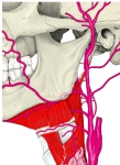 Facial artery
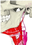 Lingual artery
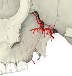 Descending palatine artery
Veins
A peritonsillar plexus drains into the pharyngeal venous plexus and the external palatine vein, and on to the facial vein and the internal jugular vein. In tonsillectomy, bleeding most often comes from the external palatine (paratonsillar) vein.
 Facial vein
 Pharyngeal venous plexus
Lymphatics
There are many. They begin in plexuses around the lymphoid nodules, pass the lateral surface of the tonsil, pierce the wall of the pharynx and enter the jugulodigastric (tonsillar) node of the deep cervical lymph nodes, below and behind the angle of the mandible near the greater horn of the hyoid bone.
Nerves
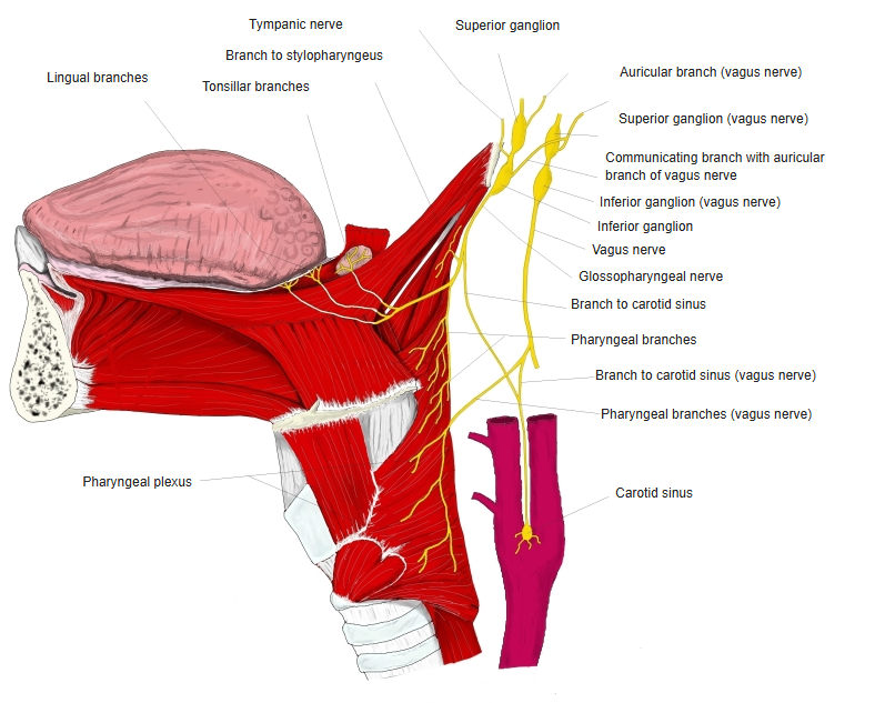
· Branches from the pharyngeal plexus (formed by branches of the glossopharyngeal and vagus nerves) form a small tonsillar plexus.
The tonsillar plexus also receives the tonsillar branches of the glossopharyngeal nerve and the lesser palatine nerves (from the maxillary nerve, through the pterygopalatine ganglion). Pain from the tonsil is often felt in the ear. The glossopharyngeal nerve runs on past the tonsil to the back third of the tongue, so it can be injured in tonsillectomy, with loss of general sensation and taste there.
Reference websites
[Wikipedia]
· Palatine tonsil, Wikipedia.
· Waldeyer's tonsillar ring, Wikipedia.
[Websites with illustrations]
· Palatine tonsil: anatomy and histology (Kenhub)
· The oral cavity (TeachMeAnatomy)
Where it is found
Related terms
- Palatoglossal arch
- Palatopharyngeal arch
- Isthmus of fauces
- Pharyngeal tonsil
- Lingual tonsil
- Oropharynx
- Soft palate
References and sources
- Further reading: Palatine tonsil, Wikipedia.
- Further reading: Waldeyer's tonsillar ring, Wikipedia.
- Federative International Programme for Anatomical Terminology (FIPAT). Terminologia Anatomica, 2nd ed. (TA2), 2019.