Alveolar septum
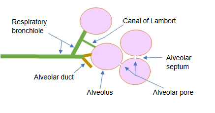
Also called the interalveolar septum (of the lung) or the alveolar wall
The interstitium (connective tissue core) of the alveolar septum contains the following:
· Capillaries · nerve fibres (some sources also list a few smooth muscle cells and lymphatic vessels; in most accounts the septum has contractile interstitial cells but no true smooth muscle, which lies only at the rims of the alveolar openings, and no lymphatic vessels)
· Interstitial tissue: fibres (collagen fibres, elastic fibres) and interstitial cells (fibroblasts, contractile cells, lipid-storing cells, macrophages and pericytes)
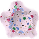 Alveolus (section)
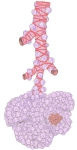 Alveolar sac
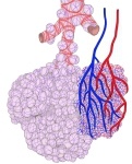 Alveoli and blood vessels
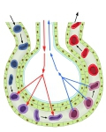 Gas exchange
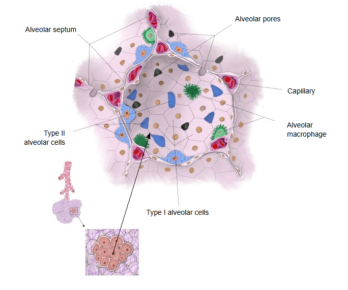
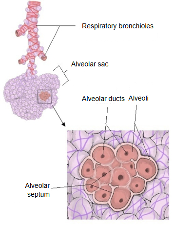
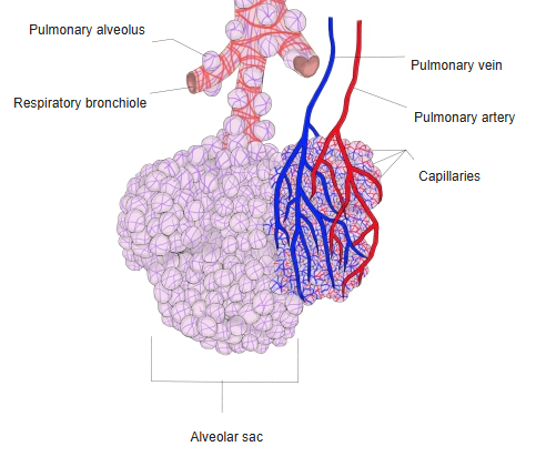
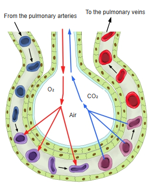
The capillary network in the septum is so dense that it has been likened to a sheet of blood, and each capillary serves the alveoli on both sides. On one side of a capillary the wall is very thin: air and blood are separated only by a type I alveolar cell, a fused basement membrane and the capillary endothelium (well under 1 µm). Gas exchange takes place mainly here.
On the other side the wall is thicker, with interstitium between the epithelium and the capillary; fluid exchange takes place mainly here. The septum has no lymphatic vessels of its own, so fluid that leaks from the capillaries drains through the interstitium to the lymphatics around the bronchioles and vessels. Neighbouring alveoli communicate through alveolar pores in the septum.
Note: do not confuse it with the interalveolar septa of the jaws, the bony plates between the tooth sockets. Sources differ on whether smooth muscle and lymphatic vessels are present in the alveolar septum itself.
Reference websites
[Wikipedia]
· Pulmonary alveolus, Wikipedia.
· Blood–air barrier, Wikipedia.
· Alveolar septum, ScienceDirect Topics.
[Websites with illustrations]
· Alveoli and capillaries (illustration) (Netter Images)

· Structure of the alveolus (Deranged Physiology)

Related terms
- Pulmonary alveoli
- Smooth muscle
- Blood
- Basement membrane
- Alveolar cavity
- Alveolar pores
- Alveolar cells
- Alveolar sac
- Alveolar lining layer
- Pulmonary circulation
- Lung
References and sources
- Further reading: Pulmonary alveolus, Wikipedia.
- Further reading: Blood–air barrier, Wikipedia.
- Federative International Programme for Anatomical Terminology (FIPAT). Terminologia Anatomica, 2nd ed. (TA2), 2019.