Hepatic lobule (Latin: lobuli hepatis)
Also called Lobule of liver or the classic lobule
- Overview
- Structure (other ways of dividing the liver tissue)
- Reference websites
- (Wikipedia, websites with illustrations)
- Related terms
Overview
· The liver parenchyma (hepatic parenchyma) is divided into many hepatic lobules by interlobular connective tissue (Glisson's sheath).
· In humans the boundaries between the lobules are not very clear, even with a magnifying glass.
The reason: in humans the Glisson's sheath between the lobules is poorly developed. In the pig the boundaries are very distinct.
⇒ A comparison of human and pig liver histology
The liver is said to consist of about 250-300 billion hepatocytes (English sources often give about 100 billion). Dividing this by the number of lobules gives
⇒ roughly 500,000 hepatocytes in one hepatic lobule (a rough calculation; estimates from the size of a lobule are lower).
Structure
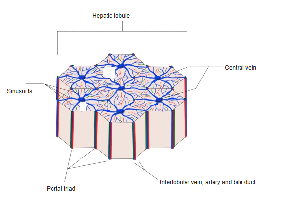
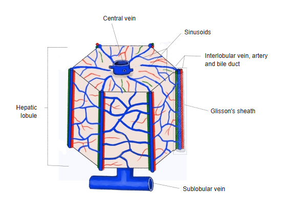
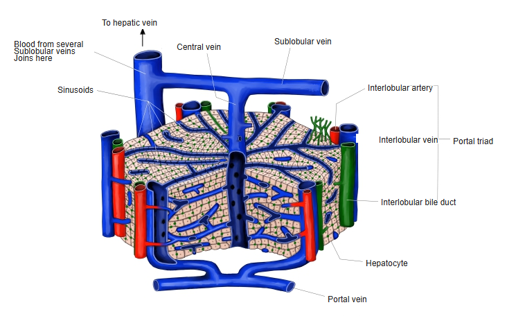
1. 1. At each corner of the prism lie an interlobular vein, an interlobular artery and a bile duct wrapped in Glisson's sheath, forming a portal triad; this area is called the portal area (portal tract).
2. 2. Blood from the interlobular arteries and veins flows through the sinusoids into the central vein, a tributary of the hepatic veins.
Bile flows the opposite way to the blood: it is made by the hepatocytes and flows toward the interlobular bile ducts.
3. 3. The hepatocytes are lined up in rows that radiate toward the central vein; these are called hepatic cell plates.
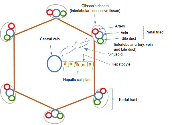
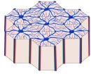 Hepatic lobules
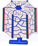 Hepatic lobule and vessels
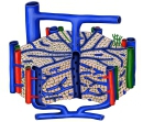 Structure of a hepatic lobule

[Other ways of dividing the liver tissue]
The hepatic lobule described above (the classic lobule) is the anatomical unit. Two other units are used to describe bile drainage and blood supply.
| Unit | Shape and centre | What it describes |
|---|---|---|
| Classic (hepatic) lobule | Hexagonal, centred on the central vein, with portal triads at the corners | The anatomical structure |
| Portal lobule | Triangular, centred on a portal triad, with three central veins at its corners | Bile drainage (bile secretion) |
| Liver acinus | Diamond-shaped, between two central veins, centred on the vessels between two portal tracts; divided into zones 1 (near the portal tract), 2 and 3 (near the central vein) | Blood supply, oxygen and metabolic zones |
Blood entering at the edge of the lobule is richer in oxygen than blood near the central vein, so the outer and inner parts of a lobule differ in function.
Reference websites
[Wikipedia]
· Lobules of liver, Wikipedia.
[Websites with illustrations]
· Microscopic anatomy of the liver (Wikimedia Commons)

· Accessory organs in digestion: the liver, pancreas and gallbladder (OpenStax Anatomy and Physiology 2e)
Related terms
- Blood
- Portal triad
- Sinusoid
- Central vein of the liver
- Interlobular veins of the liver
- Interlobular bile duct
- Glisson's sheath
- Hepatic parenchyma
References and sources
- Further reading: Lobules of liver, Wikipedia.
- Federative International Programme for Anatomical Terminology (FIPAT). Terminologia Anatomica, 2nd ed. (TA2), 2019.