Neutrophil
Also called the neutrophil granulocyte or the polymorphonuclear leukocyte
One of the three kinds of granulocytes, the white blood cells that protect the body against invaders such as viruses and bacteria. It is the most numerous white blood cell and the first to arrive at sites of bacterial infection, where it engulfs and kills microbes.
blood
cells
- Granulocytes
- Neutrophil
- Eosinophil
- Basophil
- Monocyte : ⇒ macrophage in the tissues
- Lymphocytes
- B cell : ⇒ plasma cell
- NK cell
- T cell
In more detail
Overview
One of the three kinds of granulocytes (neutrophils, eosinophils and basophils); neutrophils make up 90–95% of the granulocytes and more than 50% of all white blood cells. As they mature, the nucleus divides into lobes (segmentation), so they are also called polymorphonuclear leukocytes.
- Neutrophil : 90–95% of the granulocytes; more than 50% of the white blood cells. Also called the polymorphonuclear leukocyte.
- Eosinophil : 2–5% of the granulocytes.
- Basophil : less than 1% of the granulocytes.
Lifespan
Neutrophils live for different times in the circulating blood and in the tissues.
In the circulating blood: less than a day, about 10–12 hours by most estimates (sources differ)
In the tissues: a few days
Shape and size
Colourless and translucent. It may be almost spherical, but when it puts out pseudopodia and moves actively by amoeboid movement it loses its round shape, so it has no fixed form.
Among the white blood cells it is larger than a lymphocyte and smaller than a monocyte or a macrophage: about 12–15 µm across. The mature nucleus has 2–5 lobes joined by thin strands, and the pale cytoplasm is full of fine granules.
Where they are and how many
Neutrophils are spread throughout the body, in the circulating blood, the bone marrow and elsewhere; the body is said to hold several hundred billion of them, and about 100 billion new ones are produced each day.
| Pool | Description | |
|---|---|---|
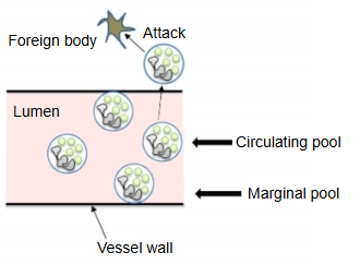
| 1 | Circulating blood · circulating pool · marginal pool | Where neutrophils are found is described with the word "pool". In the circulating blood there are two pools, the circulating pool and the marginal pool; they hold about the same number of neutrophils and exchange cells with each other. Circulating pool: neutrophils carried round the body in the bloodstream; they are the first to attack foreign matter entering the body. This is the part measured in a blood count. Marginal pool: neutrophils moving slowly (rolling) along the vessel wall; they become active when the circulating pool runs short. There are about 2,000–7,500 neutrophils per µL of blood, so an adult weighing 50 kg has about 8–30 billion neutrophils in the circulating blood. Note: figures vary between sources; 1 µL is 1/1000 mL. |
| 2 | Bone marrow pool | The bone marrow holds about 10–30 times as many neutrophils as the circulating blood, a reserve ready to be released quickly into the blood when foreign matter enters the body. |
| 3 | Others | Many are also held as a marginal pool in the spleen, the liver and elsewhere. |
Development
The diagram of the stages of development below is a simplified scheme (based on common textbook accounts).
Development of the neutrophil
Eosinophils and basophils branch off from the myeloblast stage. The neutrophil line becomes clearly recognizable from the promyelocyte and myelocyte stages.
Kinds of granules and their contents
The table below is based on common textbook accounts.
| Granule | Main contents | |
|---|---|---|
| 1 | Primary granules (azurophil granules) | Myeloperoxidase, cathepsin G, elastase, proteinase 3, defensins and others |
| 2 | Secondary granules (specific granules) | Lactoferrin, lysozyme, collagenase, cytochrome b558, cathelicidin and others |
| 3 | Tertiary granules (gelatinase granules) | Gelatinase, lysozyme, leukolysin and others |
| 4 | Secretory vesicles | Complement receptors, CD14, CD16, formyl peptide receptors |
Function
① Bacteria or other microbes enter the body.
② Macrophages and mast cells react first and release cytokines.
③ The tissue becomes inflamed.
④ Factors that attract neutrophils (chemotactic factors) are released by:
· the cytokines released by macrophages and mast cells · the inflamed tissue · the bacteria themselves · complement
Typical neutrophil chemotactic factors are:
| Factor | Notes | |
|---|---|---|
| 1 | IL-8 | Interleukin 8 (CXCL8) |
| 2 | NAP-2 | Neutrophil-activating protein 2 (CXCL7) |
| 3 | MIP-2 | Macrophage inflammatory protein 2 (CXCL2), a chemokine |
| 4 | Leukotriene B4 | Leukotrienes are eicosanoids: lipid mediators made from arachidonic acid by 5-lipoxygenase that act on the cell itself or on nearby cells (autocrine/paracrine). Leukotriene B4 attracts neutrophils (chemotaxis) and so gathers the cells needed in inflamed tissue. Leukotrienes also strongly constrict blood vessels (especially small veins) and the bronchi, and increase vascular permeability. |
| 5 | fMLP | N-formyl-methionyl-leucyl-phenylalanine: a chemotactic peptide from bacterial components |
⑤ Neutrophils sense the chemotactic factors with their receptors and start to migrate.
⑥ They gather at the site of infection.
The site of infection is usually outside the vessels. The neutrophils stick to the vessel wall, put out pseudopodia and squeeze between the endothelial cells of the vessel,
then, helped by enzymes, break through the basement membrane and leave the vessel.
⑦ They move to the inflamed area and make contact with the bacteria, which they engulf and destroy with the contents of their granules.
⇒ Scanning electron micrograph of a neutrophil engulfing anthrax bacteria (Wikimedia Commons)

Reference websites
[Wikipedia]
· Neutrophil, Wikipedia.
· Granulopoiesis, Wikipedia.
[Websites with illustrations]
· Neutrophil: anatomy, histology and function (Kenhub)
· Simplified hematopoiesis (diagram) (Wikimedia Commons)

· Neutrophil engulfing anthrax bacteria (SEM) (Wikimedia Commons)
Related terms
- White blood cell
- Granulocyte
- Blood
- Bone marrow
- Red bone marrow
- Hematopoietic stem cell
- B lymphocyte
- Basement membrane
References and sources
- Further reading: Neutrophil, Wikipedia.
- Further reading: Granulopoiesis, Wikipedia.
- Federative International Programme for Anatomical Terminology (FIPAT). Terminologia Anatomica, 2nd ed. (TA2), 2019.