Hair cell (Latin: cellula pilosa)
Also called the hair cells
· Hair cells are epithelial cells, not neurons. Each sits among supporting cells and is contacted at its base by nerve fibres of the vestibulocochlear nerve.
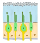
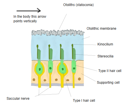
· The "hairs" are stereocilia, long microvilli arranged in rows of rising height. Vestibular hair cells also have one true cilium, the kinocilium, beside the tallest row.
· Bending the bundle toward the tallest stereocilia opens ion channels at their tips; potassium from the endolymph flows in and the cell releases transmitter onto the nerve fibres. Bending it the other way reduces the release.
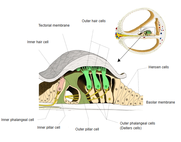
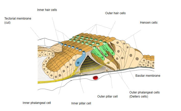
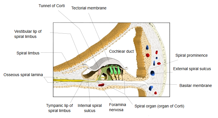
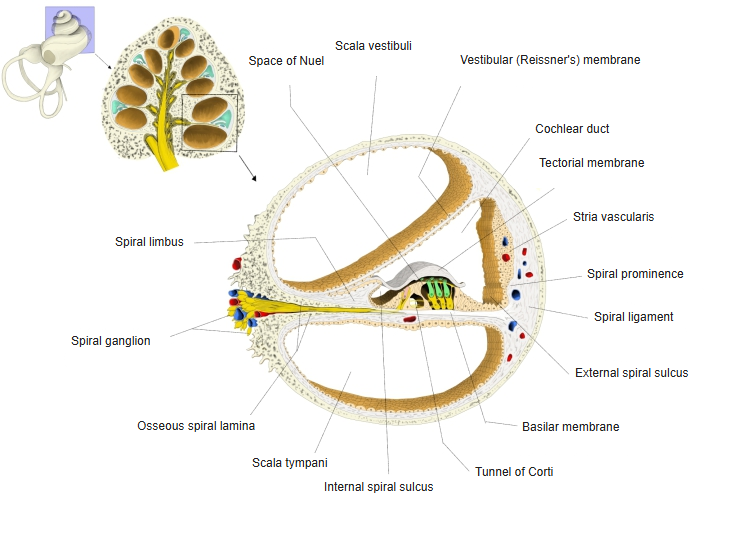
· The hair cells of the spiral organ are of two kinds: inner hair cells in one row and outer hair cells in three rows, on either side of the pillar cells; they rest on phalangeal cells (inner phalangeal cells under the inner hair cells, outer phalangeal cells under the outer hair cells).
· Inner hair cells are the main receptors for hearing; outer hair cells change their length and amplify the vibration of the basilar membrane. Mature cochlear hair cells have lost the kinocilium; the tips of their tallest stereocilia touch or lie close to the tectorial membrane.
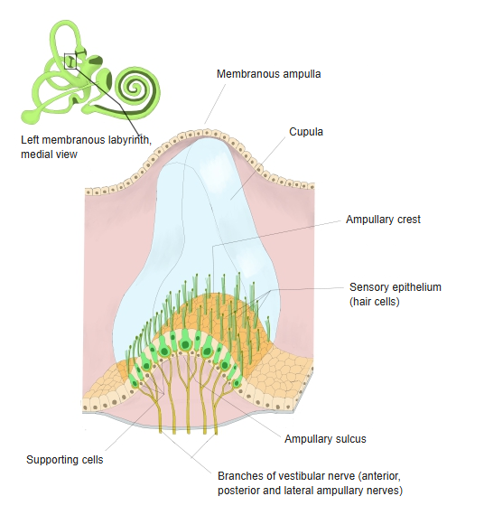
· The hair cells of the macula of the saccule and the macula of the utricle (and of the ampullary crests) are of two types: type I hair cells are flask-shaped and enclosed by a cup-like nerve ending (calyx); type II hair cells are cylindrical and receive small button-like endings.
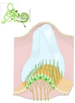
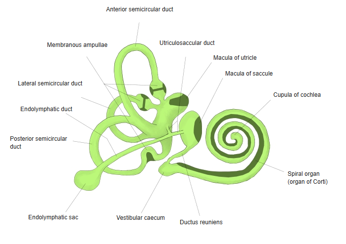
· In the maculae the hair bundles project into a gel layer carrying calcium carbonate crystals (otoliths), so the cells sense gravity and straight-line acceleration. In the ampullary crests they project into a gel cap (cupula) that is pushed by the endolymph when the head rotates.
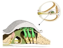 Spiral organ (section)
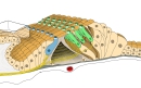 Spiral organ (tectorial membrane removed)
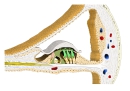 Cochlear duct
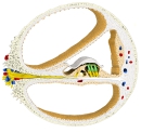 Cochlear spiral canal
 Macula of saccule and utricle
 Membranous labyrinth
 Ampullary crest
Related terms
- Inner hair cell
- Outer hair cell
- Organ of Corti
- Macula of saccule
- Macula of utricle
- Ampullary crest
- Outer phalangeal cell
References and sources
- Further reading: Hair cell, Wikipedia.
- Federative International Programme for Anatomical Terminology (FIPAT). Terminologia Anatomica, 2nd ed. (TA2), 2019.