Killer T cell
Also called the cytotoxic T cell (CTL) or CD8+ T cell
A kind of lymphocyte that protects the body by attacking body cells infected by bacteria or viruses, and cancer cells.
More details
One of the T cells (T lymphocytes); it is more often called the cytotoxic T cell (CTL). A killer T cell can attack foreign cells only after it has been activated; before activation it is called a naive CD8+ T cell. Killer T cells carry the surface marker CD8 and recognise antigen peptides shown on MHC class I molecules, which almost all nucleated cells carry, so they can find an infected cell anywhere in the body. They arise from stem cells in the bone marrow, mature in the thymus and then circulate between the blood and secondary lymphoid organs such as the lymph nodes and the spleen.
From the entry of a foreign substance to the activation of the killer T cell
Note: the steps below are a simplified outline.
| 1 | Bacteria, viruses or other foreign substances enter the body, or a normal body cell becomes cancerous; these become antigens. | 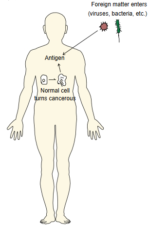 |
| 2 | An antigen-presenting cell, such as a macrophage or a dendritic cell, takes up the foreign substance (or fragments of it). | 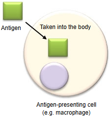 |
| 3 | It breaks the substance down into peptides, binds them to MHC class I and class II molecules and shows them on its surface (antigen presentation). | 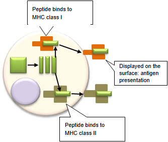 |
| 4 | When the T-cell receptor (TCR) on the surface of a helper T cell recognises "MHC class II + antigen peptide", the helper T cell is activated and produces and releases interleukin-2 (IL-2). | 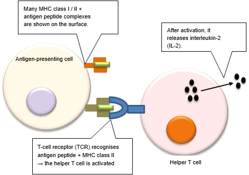 |
| 5 | The killer T cell before activation is called a naive CD8+ T cell. To be activated into a killer T cell, it needs two things, which take place at the same time: (1) the T-cell receptor on its surface recognises "MHC class I molecule + antigen peptide" on the antigen-presenting cell; (2) it receives a signal from co-stimulatory molecules. | 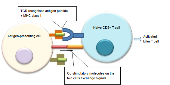 |
| 6 | Only after activation do the cells multiply: under the action of interleukin-2 from the helper T cell (and from the activated cells themselves), the activated CD8+ T cell divides again and again, giving many killer T cells that recognise the same antigen. | 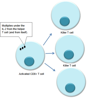 |
| 7 | The activated killer T cell releases cytotoxic substances (perforin, granzymes) and cytokines such as TNF (tumour necrosis factor), and makes the target cell undergo apoptosis. After their work, some killer T cells remain as memory killer T cells, ready to attack quickly if the same foreign substance enters again. | 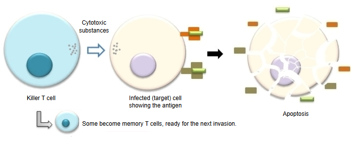 |
About co-stimulatory molecules
Co-stimulatory molecules are a group of molecules expressed on antigen-presenting cells and T cells (for example CD80/CD86 on the antigen-presenting cell and CD28 on the T cell). The co-stimulatory signal they pass decides whether the T cell becomes active and strongly affects the immune response that follows. Without it, recognition of the antigen alone does not activate the T cell. When the killer T cell then meets a target cell, it uses its antigen receptor together with the co-receptor CD8 to recognise the "antigen peptide + MHC class I" complex on the target, and attacks it.
How killer T cells destroy infected cells
Killer T cells destroy infected cells in two ways:
| 1 | Perforin/granzyme pathway | 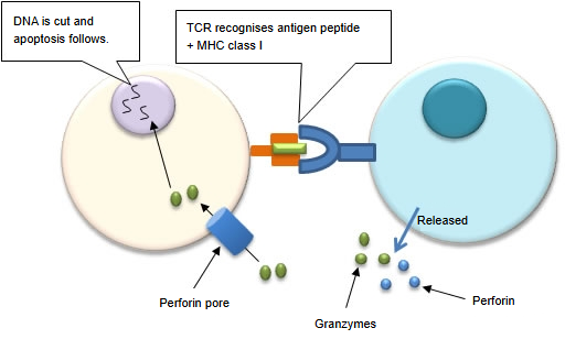 |
| When the T-cell receptor of the killer T cell recognises the antigen presentation "MHC class I molecule + antigen peptide" of an infected cell, it secretes the proteins perforin and granzymes. Perforin forms tube-shaped polymers that pierce the cell membrane of the infected cell. Granzymes enter the cell through these pores and set off its death programme: the DNA in the nucleus is cut and the cell dies by apoptosis. | ||
| 2 | Fas and Fas ligand | 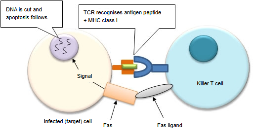 |
| When the T-cell receptor of the killer T cell recognises "MHC class I molecule + antigen peptide" on an infected cell, and the Fas ligand of the killer T cell binds the Fas molecule on the surface of the infected cell, a signal that starts apoptosis passes into the cell. The target cell dies without bursting, so its contents do not spread the infection. |
Reference websites
[Wikipedia]
· Cytotoxic T cell, Wikipedia.
Related terms
- T cell
- Helper T cell
- Apoptosis
- Lymphocyte
- White blood cell
- B lymphocyte
- Plasma cell
- Macrophage
- Dendritic cell
- Lymph node
References and sources
- Further reading: Cytotoxic T cell, Wikipedia.
- Federative International Programme for Anatomical Terminology (FIPAT). Terminologia Anatomica, 2nd ed. (TA2), 2019.