T cell
Also called T lymphocyte
Overview
cytes
- T cells : = T lymphocytes; about 60–70%. Function: cell-mediated immunity (removal of infected cells and tumour cells).
- B cells : = B lymphocytes; about 10–20%. Function: humoral immunity (production of antibodies).
- NK cells : = natural killer cells; about 10–15%. Function: non-specific (innate) immunity, such as the removal of virus-infected cells.
| Location | Group | Name | Share | Notes |
|---|---|---|---|---|
| In the peripheral blood | Granulocytes | Neutrophils | 50–75% (all granulocytes) | About 90–95% of the granulocytes |
| Eosinophils | About 2–5% of the granulocytes | |||
| Basophils | 1% or less of the granulocytes | |||
| Lymphocytes | 20–40% | Kinds: NK cells, B cells, T cells | ||
| Monocytes | 3–6% | The largest of the white blood cells | ||
| In the tissues | Dendritic cells | less than 0.1–1% | Found throughout the body, beginning with the subcutaneous tissue | |
· T cells that have differentiated and matured in the thymus are called naive T cells; they travel in the blood to the secondary lymphoid organs: the lymph nodes (mainly the paracortex), the spleen, the tonsils, the Peyer's patches and other mucosal lymphoid tissue.
· There an antigen-presenting cell shows them a fragment of an antigen (antigen presentation); the matching T cells are activated, divide and differentiate, and either act on the spot or travel through the blood to the tissues of the whole body. Under the light microscope T cells look like other small lymphocytes; they are told apart by surface markers named with CD numbers, such as CD4 and CD8.
T lymphocytes / T cells: about 60–70% of all lymphocytes; the core of acquired immunity
The cells travel from the bone marrow to the thymus, and from the thymus to the secondary lymphoid organs, in the venous blood.
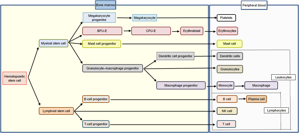
⇒ Diagram of the differentiation of hematopoietic stem cells
Development
The development of T cells in brief:
In the bone marrow
1. 1. Hematopoietic stem cells give rise to common lymphoid progenitors.
Note: hematopoietic stem cells are the source of all blood cells; common lymphoid progenitors are the source of the lymphocyte line.
2. 2. Common lymphoid progenitors become T precursor cells.
Carried in the blood to the thymus
3. 3. In the cortex of the thymus they become immature T cells and multiply.
At this stage they are "CD4⁻CD8⁻": the T-cell surface proteins (markers of the cell membrane) are not yet expressed.
4. 4. Rearrangement of the T-cell receptor (TCR)
Gene rearrangement gives each T cell its own TCR.
5. 5. Positive selection
Only cells that can recognise the body's own MHC molecules survive (in the thymic cortex).
At this point each cell becomes either CD4⁺ or CD8⁺.
6. 6. Negative selection
Cells that react strongly with the body's own antigens are removed (mainly in the thymic medulla).
This is an important step for preventing autoimmunity. Most of the developing cells die in the thymus.
7. 7. Mature T cells (naive T cells)
Only suitable T cells, which recognise the body's own MHC weakly, remain.
They leave the thymus for the peripheral lymphoid organs (lymph nodes, spleen and others). The bone marrow and the thymus are the primary lymphoid organs.
Development of T cells (simplified; the JP original notes that it may be inaccurate)
DN (double-negative) cells: CD4⁻CD8⁻, many in the region under the capsule of the cortex. DP (double-positive) cells: CD4⁺CD8⁺, about 70–80% of normal thymocytes, in the cortex; they express only about 1/10 as much TCR as mature T cells. Repertoire selection: only about 3–5% are allowed to mature.
Types
T cells are classified by the markers expressed on the surface of their cell membrane; these markers are organized by CD numbers (CD = cluster of differentiation). Each kind of cell has a different function.
| Name | Description | |
|---|---|---|
| 1 | Helper T cell (CD4) | Releases cytokines that activate other cells, such as macrophages, killer T cells and B cells. There are several subsets, including Th1 and Th2. ⇒ More |
| 2 | Killer T cell (CD8) | = cytotoxic T cell. Kills virus-infected cells and tumour cells by leading them into apoptosis (programmed cell death). ⇒ More |
| 3 | Suppressor T cell | Proposed (by a Japanese researcher) as a cell that suppresses immune reactions and brings them to an end, and studied intensively for a time; whether such a separate cell exists was later questioned. Its role is now attributed to regulatory T cells. |
| 4 | Regulatory T cell | Specialized in suppressing immunity (holding back the activation of T cells); helps prevent autoimmune disease. There are two kinds: natural ones, which arise in the thymus, and induced ones, which differentiate from naive T cells in the periphery. |
| 5 | NKT cell (natural killer T cell) | Has features of both natural killer cells and T cells. ⇒ More |
| 6 | Gamma-delta (γδ) T cell | There are two kinds of T-cell receptor (TCR): αβ and γδ. Most T cells in the peripheral blood are αβ, but γδ T cells make up a few percent. The αβ receptor recognises an antigen peptide bound to an MHC molecule, while γδ cells recognise, for example, isopentenyl pyrophosphate (IPP) and the MHC class I-related chains A/B (MICA/B). |
Memory T cells: long-lived cells that respond quickly at a second meeting with the same antigen.
Reference websites
[Wikipedia]
· T cell, Wikipedia.
· Thymocyte, Wikipedia.
Related terms
- Helper T cell
- Killer T cell
- Lymphocyte
- B lymphocyte
- Primary lymphoid organ
- Lymphoid organ
- Lymph node
- Hematopoietic stem cell
- White blood cell
References and sources
- Further reading: T cell, Wikipedia.
- Federative International Programme for Anatomical Terminology (FIPAT). Terminologia Anatomica, 2nd ed. (TA2), 2019.