Major calyx (Latin: calyx renalis major)
Plural: major calyces
Overview
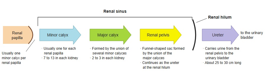
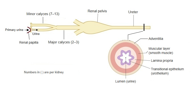
The major calyx is one of the parts of the renal sinus that form the passage of urine; several minor calyces join to form a wide tube.
There are usually two or three major calyces, which join to form the renal pelvis
· There are usually two or three major calyces, the primary branches of the renal pelvis. By position they are called the superior, middle and inferior calyces.
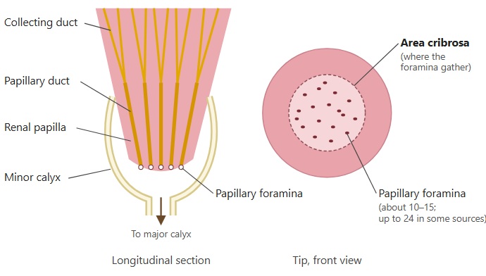
· Urine flows from the renal papilla into a minor calyx, then into a major calyx, the renal pelvis and the ureter.
- Urinary passages : minor calyx → major calyx → renal pelvis, which leaves through the renal hilum and continues as the ureter
- Other contents
- Blood vessels : branches of the renal artery and tributaries of the renal veins
- Lymphatic vessels : collect the lymph of the renal cortex and medulla
- Nerves : the renal plexus (autonomic nerves) around the renal artery, entering the sinus with its branches
- Adipose tissue : in adults it fills most of the space; at the renal hilum it is continuous with the fat around the kidney, so it is not a separate tissue
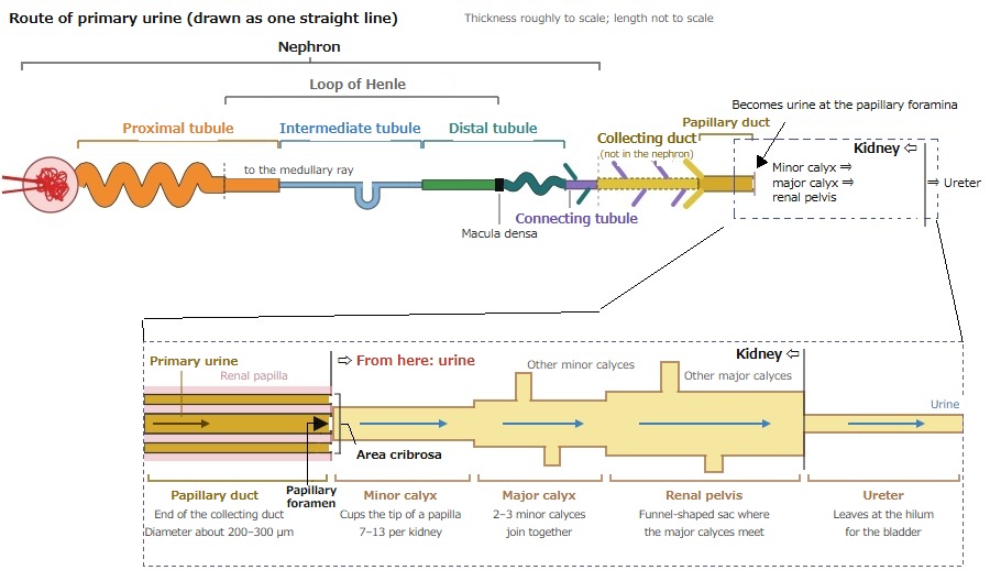
· The wall of the calyces contains smooth muscle, whose peristalsis pushes urine on into the renal pelvis and ureter.
 From the minor calyx to the ureter
 Renal papilla and papillary foramina
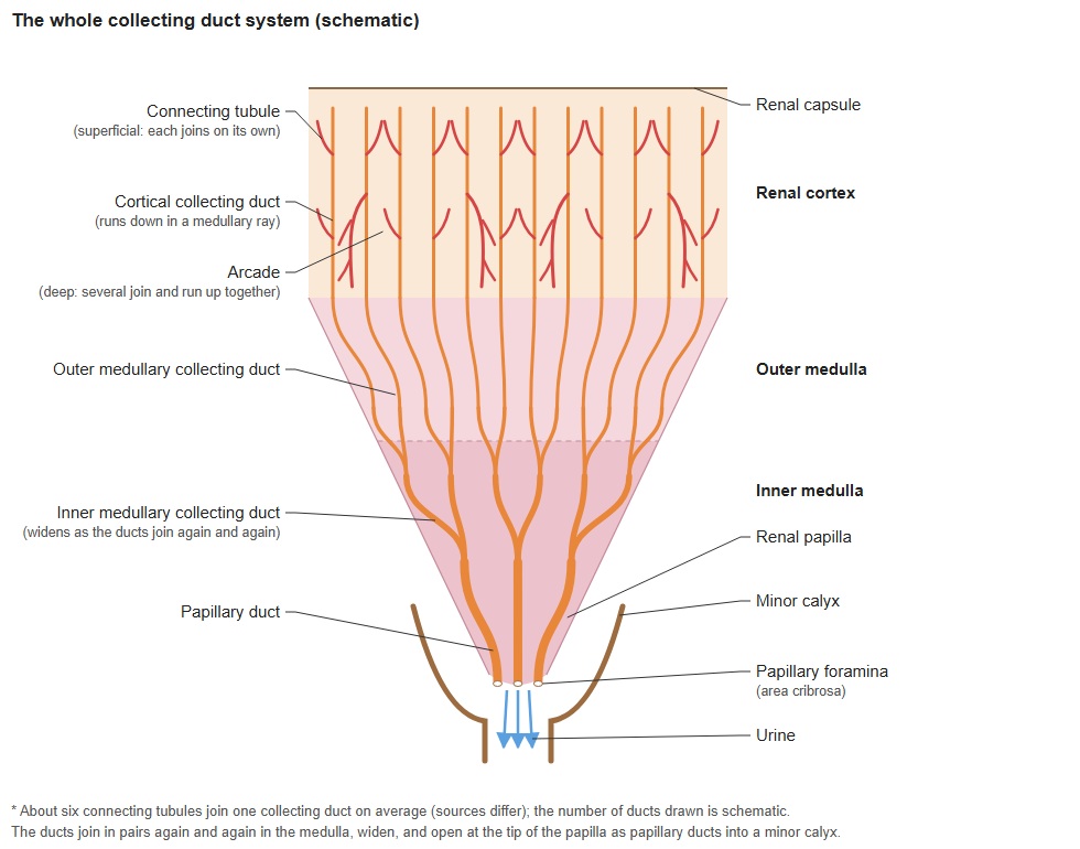
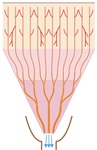 The whole collecting duct
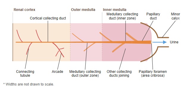
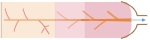 Flow through the collecting duct
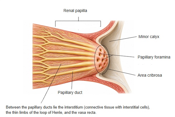
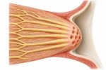 Renal papilla
Reference websites
[Wikipedia]
· Renal calyx, Wikipedia.
[Websites with illustrations]
· Hydronephrosis (ultrasound) (NYSORA)
· Gross anatomy of the kidney (Lumen Learning)
· Kidney cross-section anatomy (EBM Consult)
· Cross-section of the kidney (Encyclopaedia Britannica)

Related terms
References and sources
- Further reading: Renal calyx, Wikipedia.
- Federative International Programme for Anatomical Terminology (FIPAT). Terminologia Anatomica, 2nd ed. (TA2), 2019.