Renal sinus (Latin: sinus renalis)
Overview
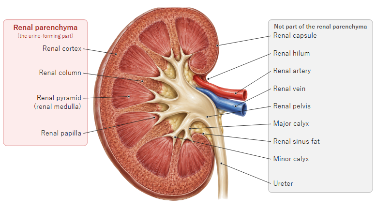
· The renal sinus is the large space inside the kidney that opens at the renal hilum on the medial border and is surrounded by the renal parenchyma.
· It contains the renal pelvis, the minor and major calyces, branches of the renal artery and renal veins, nerves, lymphatics and fat.
· The renal papillae, the tips of the renal pyramids, project into its wall, each capped by a minor calyx. Urine passes from the papillae into the minor calyces, then the major calyces and the renal pelvis, and leaves through the ureter.
· The fibrous capsule turns in at the hilum to line the sinus, and the sinus fat is continuous through the hilum with the perirenal fat.
· Sinus fat increases with age and obesity.
· On ultrasound the sinus looks brighter (more echogenic) than the parenchyma, which can make small stones hard to see. The cortex around it contains the cortical labyrinth and medullary rays.
Reference websites
[Wikipedia]
· Renal sinus, Wikipedia.
· Kidney, Wikipedia.
[Websites with illustrations]
· Excretory passageways (Digital Histology)
· Gray's Anatomy, figure 1127 (UNSW Embryology)

· Gray's Anatomy, figure 1127 (Wikimedia Commons)

Related terms
- Cortical labyrinth
- Medullary rays
- Renal parenchyma
- Kidney
- Renal pelvis
- Fibrous capsule of kidney
- Perirenal fat capsule
- Renal artery
- Ureter
References and sources
- Further reading: Renal sinus, Wikipedia.
- Further reading: Kidney, Wikipedia.
- Federative International Programme for Anatomical Terminology (FIPAT). Terminologia Anatomica, 2nd ed. (TA2), 2019.