Renal calyx (Latin: calyx renalis)
Plural: renal calyces
Overview
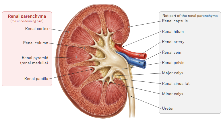
· A renal calyx is a cup-shaped tube in the renal sinus of the kidney. It receives urine from a renal papilla and passes it to the renal pelvis; it is the first part of the urinary tract. The calyces are divided into minor calyces and major calyces.
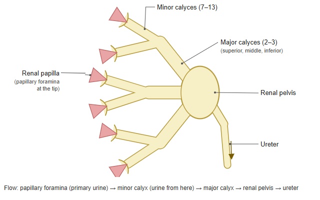
· Urine made in the kidney leaves through the many papillary foramina at the tips of the renal pyramids; the calyces are the cup-shaped sacs around the papillae that catch it.
· Each minor calyx encloses one renal papilla (there are about 7 to 13 minor calyces). Several minor calyces join to form a major calyx (2 or 3, called the superior, middle and inferior major calyces according to position). The major calyces join to form the renal pelvis.
· Urine flows in this order: papillary foramina of the area cribrosa → minor calyx → major calyx → renal pelvis → ureter.
· The inner surface of the calyces is lined by transitional epithelium (urothelium), and the wall contains smooth muscle. Peristalsis of this smooth muscle, started by pacemaker cells in the calyx wall, pushes urine on into the renal pelvis and ureter.
· A large stone that extends from the renal pelvis into the calyces is called a staghorn calculus. On imaging of the urinary tract, a normal minor calyx looks cupped at its tip because the renal papilla projects into it; when urine flow is blocked and pressure rises (hydronephrosis), this cup becomes shallow and flat.
Reference websites
[Wikipedia]
· Renal calyx, Wikipedia.
· Kidney, Wikipedia.
[Websites with illustrations]
· Renal pelvis (Kenhub)
· The kidneys (TeachMeAnatomy)
· Kidney structures (diagram) (Wikimedia Commons)

· Gray's Anatomy, fig. 1127 (Wikimedia Commons)

Related terms
References and sources
- Further reading: Renal calyx, Wikipedia.
- Further reading: Kidney, Wikipedia.
- Federative International Programme for Anatomical Terminology (FIPAT). Terminologia Anatomica, 2nd ed. (TA2), 2019.