Sphenopetrosal fissure (Latin: fissura sphenopetrosa)
Also called the sphenopetrosal suture or sphenopetrosal synchondrosis.
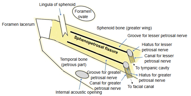
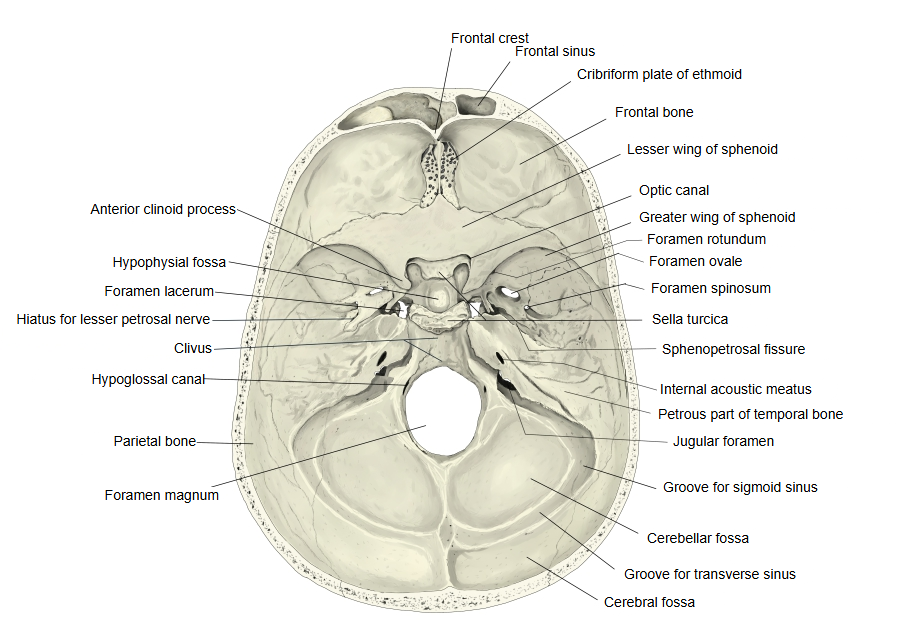
The sphenopetrosal fissure is the cleft in the middle cranial fossa between the greater wing of the sphenoid bone and the petrous part of the temporal bone.
It runs backwards and laterally from the foramen lacerum; beside it, on the front surface of the petrous part, lie the grooves and hiatuses for two nerves, the greater and lesser petrosal nerves (groove / hiatus for greater petrosal nerve, groove / hiatus for lesser petrosal nerve).
The foramen lacerum is formed between the apex of the petrous part of the temporal bone and the sphenoid bone.
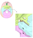 Foramen lacerum → sphenopetrosal fissure
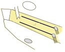 Sphenopetrosal fissure (diagram)
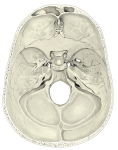 Internal cranial base
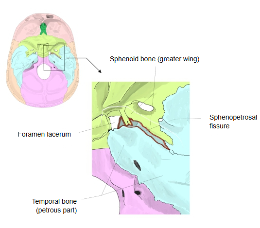
· In life the fissure is filled with cartilage and fibrous tissue. On the outer surface of the skull base it continues as the sulcus for the auditory tube, which holds the cartilaginous part of the tube, and the lesser petrosal nerve may leave the skull through it.
Reference websites
[Wikipedia]
· Sphenopetrosal fissure, Wikipedia.
· Middle cranial fossa, Wikipedia.
[Websites with illustrations]
· Middle cranial fossa: boundaries and foramina (TeachMeAnatomy)
Related terms
- Middle cranial fossa
- Greater wing
- Petrous part
- Foramen lacerum
- Apex of petrous part
- Sulcus for auditory tube
- Groove for lesser petrosal nerve
References and sources
- Further reading: Sphenopetrosal fissure, Wikipedia.
- Federative International Programme for Anatomical Terminology (FIPAT). Terminologia Anatomica, 2nd ed. (TA2), 2019.