Groove for lesser petrosal nerve (Latin: sulcus nervi petrosi minoris)
- Squamous part : the part that takes up the middle and the whole upper part of the bone; it has a temporal surface and a cerebral surface.
- Petrous part
- Mastoid part : below the squamous part (from the mastoid process to the parietal notch).
- Pyramid : a three-sided pyramid running forwards and medially from the boundary of the squamous and mastoid parts; it has an apex, surfaces and borders.
- Apex : points towards the foramen lacerum; the internal opening of the carotid canal opens here.
- Anterior surface : = cerebral surface: trigeminal impression, arcuate eminence, grooves for the greater and lesser petrosal nerves.
- Posterior surface : = cerebellar surface: internal acoustic opening, subarcuate fossa, external aperture of the vestibular aqueduct.
- Inferior surface : = basal surface: styloid process, jugular fossa, groove for the mastoid canaliculus, external aperture of the cochlear canaliculus, external opening of the carotid canal, petrous fossula, inferior opening of the tympanic canaliculus.
- Tympanic part : the smallest part: a thin, half-tube-shaped plate of bone that forms the front and lower walls of the external acoustic meatus.
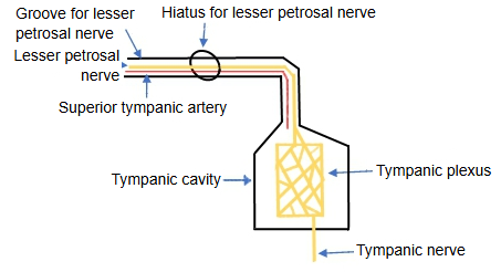
· The lesser petrosal nerve comes from the glossopharyngeal nerve (cranial nerve IX). Its tympanic nerve, from the inferior ganglion, enters the tympanic cavity through the tympanic canaliculus and forms the tympanic plexus on the promontory. The lesser petrosal nerve leaves the plexus, passes upwards through a short canal (the canal for the lesser petrosal nerve) and the hiatus, and runs in this groove. It carries parasympathetic fibres that, after relay in the otic ganglion, supply the parotid gland.
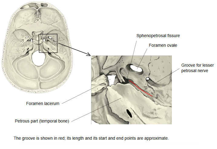
· From the hiatus the groove runs forwards and medially, just lateral to and parallel with the groove for the greater petrosal nerve, and ends near the back end of the sphenopetrosal fissure, pointing towards the foramen ovale. The nerve leaves the skull through the foramen ovale (or a small nearby foramen) to reach the otic ganglion. In the skull view below, the red line marking the groove is approximate.
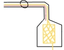 Lesser petrosal nerve leaving the tympanic cavity (diagram)
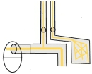 Greater and lesser petrosal grooves (diagram)
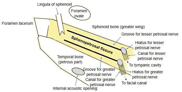
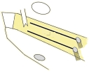 Sphenopetrosal fissure (diagram)
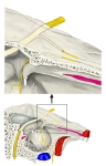 Right tympanic cavity, medial wall (oblique section)
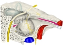 Right tympanic cavity, medial wall
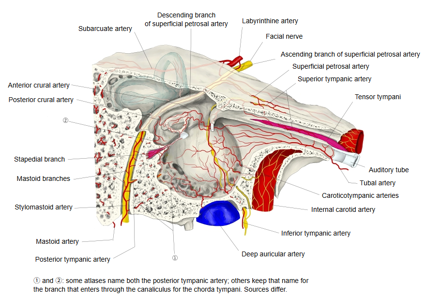
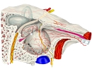 Arteries of the tympanic cavity
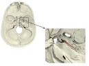 Internal cranial base
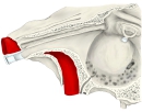 Left tympanic cavity (anterolateral view)
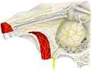 Glossopharyngeal branches in the tympanic cavity
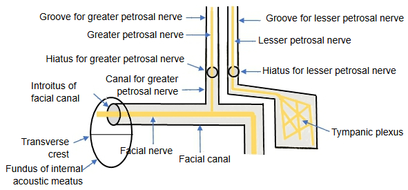
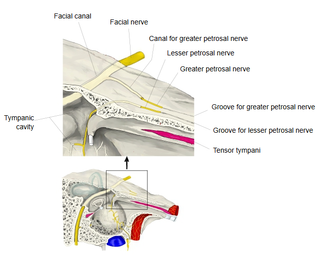
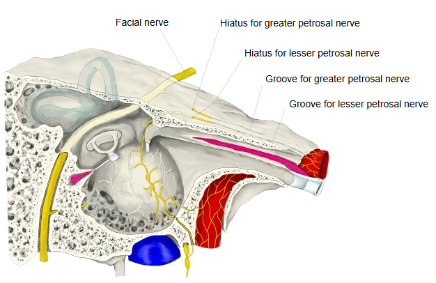
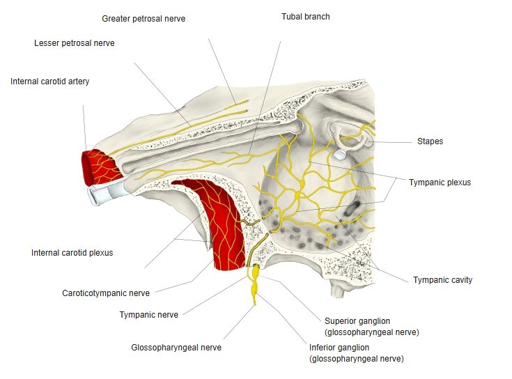
Behind the trigeminal impression on the anterior surface of the petrous part are two small grooves that run almost parallel to its long axis. The medial one is the groove for the greater petrosal nerve, the path of the greater petrosal nerve; at its back end it leads through the hiatus for the greater petrosal nerve into the facial canal inside the bone. The lateral one is the groove for the lesser petrosal nerve, which carries that nerve; at its back end it enters the bone at the hiatus for the lesser petrosal nerve and continues into the canal for the lesser petrosal nerve.
The sphenopetrosal fissure runs backwards and laterally from the foramen lacerum, between the greater wing of the sphenoid and the petrous part. The front end of the groove for the greater petrosal nerve, which lies medial to and behind the fissure, reaches the foramen lacerum, while the groove for the lesser petrosal nerve runs from the back end of the fissure towards the foramen ovale.
Reference websites
[Wikipedia]
· Lesser petrosal nerve, Wikipedia.
· Petrous part of the temporal bone, Wikipedia.
[Websites with illustrations]
· The glossopharyngeal nerve (CN IX) (TeachMeAnatomy)
· Left temporal bone, inner surface (Gray's Anatomy, fig. 138) (Wikimedia Commons)

Related terms
- Canal for greater petrosal nerve
- Groove for greater petrosal nerve
- Hiatus for lesser petrosal nerve
- Canal for lesser petrosal nerve
- Lesser petrosal nerve
- Petrous part
- Foramen ovale
- Otic ganglion
- Tympanic plexus
- Superior opening of tympanic canaliculus
- Temporal bone
References and sources
- Further reading: Lesser petrosal nerve, Wikipedia.
- Further reading: Petrous part of the temporal bone, Wikipedia.
- Federative International Programme for Anatomical Terminology (FIPAT). Terminologia Anatomica, 2nd ed. (TA2), 2019.