Sphincter pupillae (Latin: musculus sphincter pupillae)
Also called the pupillary sphincter, iris sphincter muscle
The sphincter pupillae is a sphincter muscle:
ocular
muscles
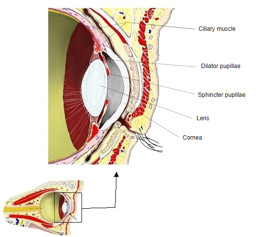
- Ciliary muscle : surrounds the lens and adjusts its thickness.
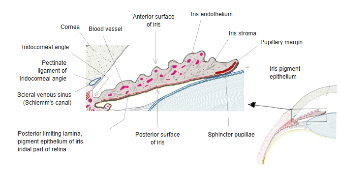
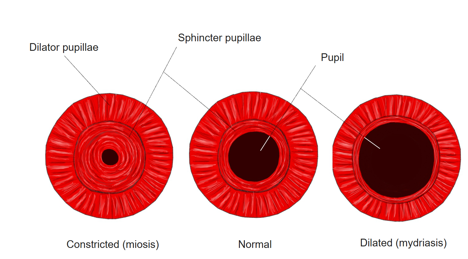
- Sphincter pupillae : runs in a ring in the iris and makes the pupil smaller. One of the muscles of the iris.
- Dilator pupillae : runs radially in the outer part of the iris and makes the pupil larger. One of the muscles of the iris.
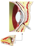 Front of the orbit (section)
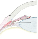 Iris (section)
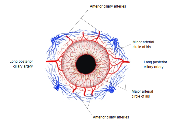
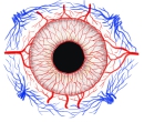 Vessels of the iris
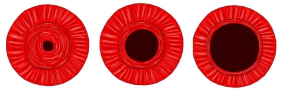 Size of the pupil
- Nucleus of oculomotor nerve : motor fibres. Superior branch: superior rectus and levator palpebrae superioris. Inferior branch: medial rectus, inferior rectus and inferior oblique.
- Accessory nucleus (Edinger-Westphal nucleus) : parasympathetic fibres: inferior branch → ciliary ganglion → short ciliary nerves → sphincter pupillae and ciliary muscle.
- Course : leaves the midbrain at the medial side of the cerebral peduncle and enters the orbit through the superior orbital fissure; it divides into the superior and inferior branches in the front part of the cavernous sinus, just before or as it passes through the fissure.
Some sources divide the muscles of the eye broadly into intra-ocular and extra-ocular muscles,
but others do not count the intra-ocular muscles among the "ocular muscles" at all, probably because they are smooth muscle.
Below are the ocular muscles in that narrower sense; some sources also leave out levator palpebrae superioris, the last item.
- Recti : superior rectus, inferior rectus, medial rectus, lateral rectus: move the eyeball.
- Obliques : superior oblique, inferior oblique: move the eyeball.
- Levator palpebrae superioris : raises the upper eyelid.
- Nerve supply : oculomotor nerve: superior, inferior and medial rectus, inferior oblique, levator palpebrae superioris; trochlear nerve: superior oblique; abducens nerve: lateral rectus.
The next classification divides the ocular muscles broadly into the extra-ocular muscles (striated muscle) and the intra-ocular muscles (smooth muscle).
muscles (9)
- Extra-ocular muscles (6)
- Recti : superior rectus, inferior rectus, medial rectus, lateral rectus.
- Obliques : superior oblique, inferior oblique.
- Intra-ocular muscles (3) : ciliary muscle, sphincter pupillae, dilator pupillae.
- Other muscles in the orbit : levator palpebrae superioris, orbitalis, superior tarsal muscle, inferior tarsal muscle.
Useful sites
Reference websites
[Wikipedia]
· Iris sphincter muscle, Wikipedia.
· Pupillary light reflex, Wikipedia.
[Websites with illustrations]
· Sphincter and dilator pupillae (SlideShare (image))

Related terms
- Muscles of iris
- Intra-ocular muscles
- Ciliary muscle
- Pupillary margin of iris
- Stroma of iris
- Smooth muscle
- Extra-ocular muscles
References and sources
- Further reading: Iris sphincter muscle, Wikipedia.
- Further reading: Pupillary light reflex, Wikipedia.
- Federative International Programme for Anatomical Terminology (FIPAT). Terminologia Anatomica, 2nd ed. (TA2), 2019.